• 文献检索
  • 文档翻译
  • 深度研究
  • 学术资讯
  • Suppr Zotero 插件Zotero 插件
  • 邀请有礼
  • 套餐&价格
  • 历史记录
应用&插件
Suppr Zotero 插件Zotero 插件浏览器插件Mac 客户端Windows 客户端微信小程序
定价
高级版会员购买积分包购买API积分包
服务
文献检索文档翻译深度研究API 文档MCP 服务
关于我们
关于 Suppr公司介绍联系我们用户协议隐私条款
关注我们

Suppr 超能文献

核心技术专利:CN118964589B侵权必究
粤ICP备2023148730 号-1Suppr @ 2026

文献检索

告别复杂PubMed语法,用中文像聊天一样搜索,搜遍4000万医学文献。AI智能推荐,让科研检索更轻松。

立即免费搜索

文件翻译

保留排版,准确专业,支持PDF/Word/PPT等文件格式,支持 12+语言互译。

免费翻译文档

深度研究

AI帮你快速写综述,25分钟生成高质量综述,智能提取关键信息,辅助科研写作。

立即免费体验

喷砂酸蚀钛牙种植体表面的成体干细胞在定向分化、衰老及生物安全性方面的特性

Adult stem cells properties in terms of commitment, aging and biological safety of grit-blasted and Acid-etched ti dental implants surfaces.

作者信息

Gardin Chiara, Ferroni Letizia, Bressan Eriberto, Calvo-Guirado José L, Degidi Marco, Piattelli Adriano, Zavan Barbara

机构信息

Department of Biomedical Sciences, University of Padua, Padua, Italy.

Department of Neurosciences, University of Padua, Padua, Italy.

出版信息

Int J Mol Cell Med. 2014 Fall;3(4):225-36.

PMID:25635249
原文链接:https://pmc.ncbi.nlm.nih.gov/articles/PMC4293610/
Abstract

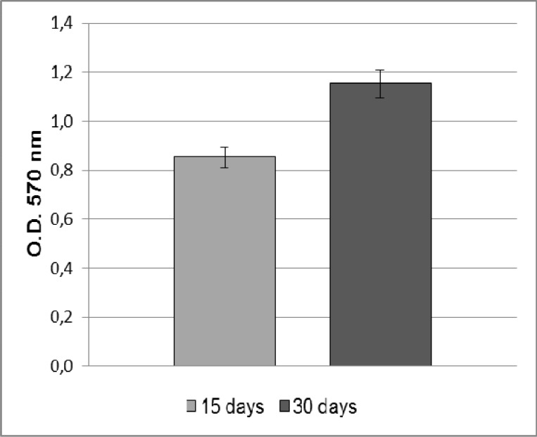
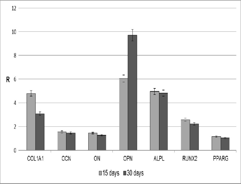
Titanium (Ti) is one of the most widely used biomaterials for manufacturing dental implants. The implant surface properties strongly influence osseointegration. The aim of the present study was to in vitro investigate the characteristics of Ti dental implants in terms of mutagenicity, hemocompatibility, biocompatibility, osteoinductivity and biological safety. The Ames test was used to test the mutagenicity of the Ti dental implants, and the hemolysis assay for evaluating their hemocompatibility. Human adipose - derived stem cells (ADSCs) were then seeded onto these implants in order to evaluate their cytotoxicity. Gene expression analyzing with real-time PCR was carried out to investigate the osteoinductivity of the biomaterials. Finally, the genetic stability of the cells cultured onto dental implants was determined by karyotyping. Our results demonstrated that Ti dental implants are not mutagenic, do not cause hemolysis, and are biocompatible. The MTT assay revealed that ADSCs, seeded on Ti dental implants, proliferate up to 30 days in culture. Moreover, ADSCs loaded on Ti dental implants show a substantial expression of some osteoblast specific markers, such as COL1A1, OPN, ALPL, and RUNX2, as well as chromosomal stability after 30 days of culture in a medium without osteogenic factors. In conclusion, the grit-blasted and acid-etched treatment seems to favor the adhesion and proliferation of ADSCs and improve the osteoinductivity of Ti dental implant surfaces.

摘要

钛(Ti)是制造牙种植体应用最广泛的生物材料之一。种植体表面特性对骨结合有很大影响。本研究的目的是在体外研究钛牙种植体在致突变性、血液相容性、生物相容性、骨诱导性和生物安全性方面的特性。采用艾姆斯试验检测钛牙种植体的致突变性,采用溶血试验评估其血液相容性。然后将人脂肪来源干细胞(ADSCs)接种到这些种植体上,以评估其细胞毒性。通过实时PCR进行基因表达分析,以研究生物材料的骨诱导性。最后,通过核型分析确定接种在牙种植体上的细胞的遗传稳定性。我们的结果表明,钛牙种植体无致突变性,不引起溶血,具有生物相容性。MTT试验显示,接种在钛牙种植体上的ADSCs在培养30天内增殖。此外,接种在钛牙种植体上的ADSCs在无成骨因子的培养基中培养30天后,显示出一些成骨细胞特异性标志物的大量表达,如COL1A1、OPN、ALPL和RUNX2,以及染色体稳定性。总之,喷砂和酸蚀处理似乎有利于ADSCs的黏附和增殖,并改善钛牙种植体表面的骨诱导性。

https://cdn.ncbi.nlm.nih.gov/pmc/blobs/2b8c/4293610/724843085874/ijmcm-3-225-g004.jpg
https://cdn.ncbi.nlm.nih.gov/pmc/blobs/2b8c/4293610/b71fdca79c19/ijmcm-3-225-g001.jpg
https://cdn.ncbi.nlm.nih.gov/pmc/blobs/2b8c/4293610/32e1e296cd40/ijmcm-3-225-g002.jpg
https://cdn.ncbi.nlm.nih.gov/pmc/blobs/2b8c/4293610/0da0f818d4db/ijmcm-3-225-g003.jpg
https://cdn.ncbi.nlm.nih.gov/pmc/blobs/2b8c/4293610/724843085874/ijmcm-3-225-g004.jpg

相似文献

1
Adult stem cells properties in terms of commitment, aging and biological safety of grit-blasted and Acid-etched ti dental implants surfaces.喷砂酸蚀钛牙种植体表面的成体干细胞在定向分化、衰老及生物安全性方面的特性
Int J Mol Cell Med. 2014 Fall;3(4):225-36.
2
Non-Washed Resorbable Blasting Media (NWRBM) on Titanium Surfaces could Enhance Osteogenic Properties of MSCs through Increase of miRNA-196a And VCAM1.钛表面的非冲洗可吸收爆破介质(NWRBM)可通过增加 miRNA-196a 和 VCAM1 来增强间充质干细胞的成骨特性。
Stem Cell Rev Rep. 2016 Oct;12(5):543-552. doi: 10.1007/s12015-016-9669-1.
3
Influence of implant surface topography on bone-regenerative potential and mechanical retention in the human maxilla and mandible.种植体表面形貌对人类上颌骨和下颌骨骨再生潜能及机械固位的影响。
Am J Dent. 2014 Jun;27(3):171-6.
4
Osteoblastic cell behaviour on modified titanium surfaces.成骨细胞在改性钛表面的行为。
Micron. 2018 Feb;105:55-63. doi: 10.1016/j.micron.2017.11.010. Epub 2017 Nov 22.
5
Novel hydrophilic nanostructured microtexture on direct metal laser sintered Ti-6Al-4V surfaces enhances osteoblast response in vitro and osseointegration in a rabbit model.直接金属激光烧结钛-6铝-4钒表面的新型亲水性纳米结构微纹理增强了体外成骨细胞反应及兔模型中的骨整合。
J Biomed Mater Res A. 2016 Aug;104(8):2086-98. doi: 10.1002/jbm.a.35739. Epub 2016 May 4.
6
Effect of surface alkali-based treatment of titanium implants on ability to promote in vitro mineralization and in vivo bone formation.钛种植体表面碱处理对体外矿化及体内骨形成促进能力的影响。
Acta Biomater. 2017 Jul 15;57:511-523. doi: 10.1016/j.actbio.2017.05.016. Epub 2017 May 9.
7
Histomorphometric analysis of the osseointegration of four different implant surfaces in the femoral epiphyses of rabbits.兔股骨骨骺中四种不同种植体表面骨结合的组织形态计量学分析。
Clin Oral Implants Res. 2008 Nov;19(11):1103-10. doi: 10.1111/j.1600-0501.2008.01547.x.
8
Biological responses of human bone mesenchymal stem cells to Ti and TiZr implant materials.人骨髓间充质干细胞对 Ti 和 TiZr 种植材料的生物学反应。
Clin Implant Dent Relat Res. 2019 Aug;21(4):550-564. doi: 10.1111/cid.12756. Epub 2019 Apr 22.
9
Titanium dental implants surface-immobilized with gold nanoparticles as osteoinductive agents for rapid osseointegration.表面固定有金纳米颗粒作为骨诱导剂的钛牙科植入物,用于快速骨整合。
J Colloid Interface Sci. 2016 May 1;469:129-137. doi: 10.1016/j.jcis.2016.02.022. Epub 2016 Feb 6.
10
The cytocompatibility and osseointegration of the Ti implants with XPEED® surfaces.具有 XPEED® 表面的钛植入物的细胞相容性和骨整合性。
Clin Oral Implants Res. 2012 Nov;23(11):1283-9. doi: 10.1111/j.1600-0501.2011.02304.x. Epub 2011 Oct 21.

引用本文的文献

1
Dentin Particulate for Bone Regeneration: An In Vitro Study.牙本质颗粒在骨再生中的应用:一项体外研究。
Int J Mol Sci. 2022 Aug 18;23(16):9283. doi: 10.3390/ijms23169283.
2
Exosomes Derived from Dental Pulp Stem Cells Show Different Angiogenic and Osteogenic Properties in Relation to the Age of the Donor.来自牙髓干细胞的外泌体在与供体年龄相关方面表现出不同的血管生成和成骨特性。
Pharmaceutics. 2022 Apr 21;14(5):908. doi: 10.3390/pharmaceutics14050908.
3
Nanostructured Modifications of Titanium Surfaces Improve Vascular Regenerative Properties of Exosomes Derived from Mesenchymal Stem Cells: Preliminary In Vitro Results.

本文引用的文献

1
Cell therapy for bone repair.用于骨修复的细胞疗法。
Orthop Traumatol Surg Res. 2014 Feb;100(1 Suppl):S107-12. doi: 10.1016/j.otsr.2013.11.010. Epub 2014 Jan 7.
2
Influence of acid-etching after grit-blasted on osseointegration of titanium dental implants: in vitro and in vivo studies.酸蚀后喷砂对钛牙科种植体骨整合影响的体外与体内研究。
J Mater Sci Mater Med. 2013 Aug;24(8):2047-55. doi: 10.1007/s10856-013-4935-0. Epub 2013 Apr 27.
3
Cryopreservation does not alter karyotype, multipotency, or NANOG/SOX2 gene expression of amniotic fluid mesenchymal stem cells.
钛表面的纳米结构修饰改善间充质干细胞来源外泌体的血管再生特性:初步体外研究结果
Nanomaterials (Basel). 2021 Dec 20;11(12):3452. doi: 10.3390/nano11123452.
4
Metal Nanoparticles Released from Dental Implant Surfaces: Potential Contribution to Chronic Inflammation and Peri-Implant Bone Loss.从牙种植体表面释放的金属纳米颗粒:对慢性炎症和种植体周围骨质流失的潜在影响
Materials (Basel). 2019 Jun 25;12(12):2036. doi: 10.3390/ma12122036.
5
Laser-modified titanium surfaces enhance the osteogenic differentiation of human mesenchymal stem cells.激光改性钛表面增强了人骨髓间充质干细胞的成骨分化。
Stem Cell Res Ther. 2017 Nov 28;8(1):269. doi: 10.1186/s13287-017-0717-9.
6
Non-Washed Resorbable Blasting Media (NWRBM) on Titanium Surfaces could Enhance Osteogenic Properties of MSCs through Increase of miRNA-196a And VCAM1.钛表面的非冲洗可吸收爆破介质(NWRBM)可通过增加 miRNA-196a 和 VCAM1 来增强间充质干细胞的成骨特性。
Stem Cell Rev Rep. 2016 Oct;12(5):543-552. doi: 10.1007/s12015-016-9669-1.
7
Adipose-Derived Stem Cells as a Tool for Dental Implant Osseointegration: an Experimental Study in the Dog.脂肪源性干细胞作为牙种植体骨结合的一种工具:犬类实验研究
Int J Mol Cell Med. 2015 Fall;4(4):197-208.
冷冻保存不会改变羊水间充质干细胞的核型、多能性或NANOG/SOX2基因表达。
Genet Mol Res. 2012 Apr 19;11(2):1002-12. doi: 10.4238/2012.April.19.5.
4
Mesenchymal stem cells and their challenges for bone regeneration and osseointegration.间充质干细胞及其在骨再生和骨整合中的挑战。
J Periodontol. 2012 May;83(5):547-50. doi: 10.1902/jop.2011.110384. Epub 2011 Sep 26.
5
Role of ox-PAPCs in the differentiation of mesenchymal stem cells (MSCs) and Runx2 and PPARγ2 expression in MSCs-like of osteoporotic patients.骨桥蛋白阳性细胞在骨质疏松患者骨髓间充质干细胞样细胞分化中的作用及对 Runx2 和 PPARγ2 表达的影响。
PLoS One. 2011;6(6):e20363. doi: 10.1371/journal.pone.0020363. Epub 2011 Jun 3.
6
In vitro concurrent endothelial and osteogenic commitment of adipose-derived stem cells and their genomical analyses through comparative genomic hybridization array: novel strategies to increase the successful engraftment of tissue-engineered bone grafts.体外同时诱导脂肪干细胞向血管内皮细胞和成骨细胞分化及其比较基因组杂交分析:提高组织工程骨移植物成功植入的新策略。
Stem Cells Dev. 2012 Mar 20;21(5):767-77. doi: 10.1089/scd.2011.0147. Epub 2011 Jun 10.
7
In vitro cell performance on hydroxyapatite particles/poly(L-lactic acid) nanofibrous scaffolds with an excellent particle along nanofiber orientation.在具有优异颗粒沿纳米纤维取向的羟基磷灰石颗粒/聚(L-乳酸)纳米纤维支架上的体外细胞性能。
Acta Biomater. 2011 Jun;7(6):2585-92. doi: 10.1016/j.actbio.2011.02.021. Epub 2011 Feb 17.
8
Melatonin inhibits adipogenesis and enhances osteogenesis of human mesenchymal stem cells by suppressing PPARγ expression and enhancing Runx2 expression.褪黑素通过抑制过氧化物酶体增殖物激活受体 γ(PPARγ)的表达和增强 runt 相关转录因子 2(Runx2)的表达来抑制人骨髓间充质干细胞的成脂分化,促进其成骨分化。
J Pineal Res. 2010 Nov;49(4):364-72. doi: 10.1111/j.1600-079X.2010.00803.x. Epub 2010 Aug 24.
9
Titanium oral implants: surface characteristics, interface biology and clinical outcome.钛质口腔种植体:表面特性、界面生物学与临床结果。
J R Soc Interface. 2010 Oct 6;7 Suppl 5(Suppl 5):S515-27. doi: 10.1098/rsif.2010.0118.focus. Epub 2010 Jun 30.
10
Regulation of osteoblast differentiation by Runx2.Runx2 对成骨细胞分化的调控。
Adv Exp Med Biol. 2010;658:43-9. doi: 10.1007/978-1-4419-1050-9_5.