Min Seiko, Liu Yi, Tang Jianxia, Xie Yilin, Xiong Jimin, You Hyung-Keun, Zadeh Homayoun H
Laboratory for Immunoregulation and Tissue Engineering, Ostrow School of Dentistry, University of Southern California, Los Angeles, CA, USA.
Laboratory of Tissue Regeneration and Immunology and Department of Periodontics, Beijing Key Laboratory of Tooth Regeneration and Function Reconstruction, Capital Medical University School of Stomatology, Beijing, China.
Clin Oral Implants Res. 2016 Jan;27(1):97-105. doi: 10.1111/clr.12521. Epub 2015 Feb 6.
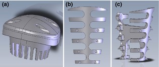
This study sought to investigate dimensional changes to the alveolar bone following extraction and application of novel devices used for obturation of socket orifice (socket cap) and space maintenance in sockets with facial dehiscence (socket cage).
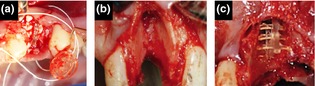
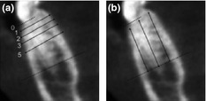
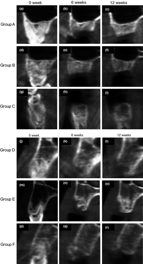
Six Macaca fascicularis had six teeth each removed according to the following intervention groups (groups A-C intact alveolar bone; D-E facial dehiscence): negative control (A); socket obturated with cap (B); filled with anorganic bovine bone mineral (ABBM) + socket cap (C); dehiscence negative control (D); socket cap + socket cage (E); ABBM + socket cap + socket cage (F). Serial CBCT scans at preoperatively, 6 and 12 weeks following intervention were compared to quantify linear alveolar bone alterations.
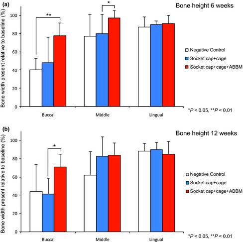
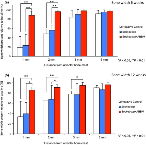
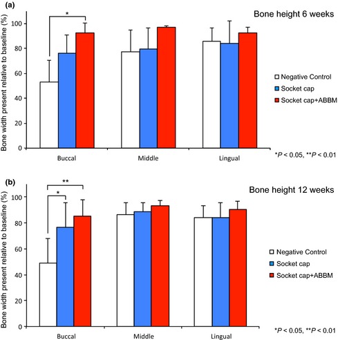
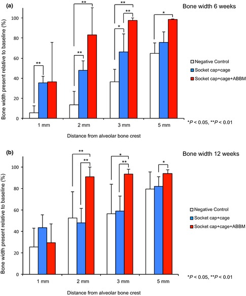
Without therapeutic intervention, intact sockets exhibited significant reduction in width at the crestal 2 mm of the ridge crest within 6 weeks. Compared with the negative control sites which lost up to 52% of crestal bone width, sites treated with socket cap + ABBM lost at most 4% of bone width at the crestal 2 mm. Similar results were seen in the dehiscence groups, with the combination of socket cap + socket cage + ABBM maintaining the greatest socket width and height dimensions.
Results from the current non-human primate study suggest that the socket cap and socket cage devices, when used in conjunction with xenograft proved effective in minimizing post-extraction socket width loss and height seen in both intact sockets and sockets with facial dehiscence defects.
本研究旨在调查拔牙后牙槽骨的尺寸变化,以及新型装置(用于封闭牙槽窝口的装置(牙槽窝帽)和用于维持存在面部骨缺损的牙槽窝空间的装置(牙槽窝笼))的应用效果。
选用6只食蟹猴,每只猴拔除6颗牙齿,分为以下干预组(A - C组为完整牙槽骨;D - E组为面部骨缺损):阴性对照组(A);用牙槽窝帽封闭牙槽窝组(B);填充无机牛骨矿物质(ABBM)+牙槽窝帽组(C);骨缺损阴性对照组(D);牙槽窝帽+牙槽窝笼组(E);ABBM+牙槽窝帽+牙槽窝笼组(F)。比较术前、干预后6周和12周的系列CBCT扫描结果,以量化牙槽骨的线性改变。
未经治疗干预时,完整牙槽窝在6周内牙槽嵴顶2mm处宽度显著减小。与阴性对照部位牙槽嵴顶骨宽度最多损失52%相比,用牙槽窝帽+ABBM治疗的部位在牙槽嵴顶2mm处骨宽度最多损失4%。在骨缺损组也观察到类似结果,牙槽窝帽+牙槽窝笼+ABBM组合能最大程度维持牙槽窝的宽度和高度尺寸。
当前非人灵长类动物研究结果表明,牙槽窝帽和牙槽窝笼装置与异种移植物联合使用时,在减少完整牙槽窝以及存在面部骨缺损的牙槽窝拔牙后牙槽窝宽度损失和高度方面证明是有效的。