Wu Xin-Bao, Wang Jun-Qiang, Zhao Chun-Peng, Sun Xu, Shi Yin, Zhang Zi-An, Li Yu-Neng, Wang Man-Yi
Department of Orthopaedic Trauma, Beijing Jishuitan Hospital; Laboratory of Bone Tissue Engineering, Beijing Research Institute of Traumatology and Orthopaedics, Beijing 100035, China.
Chin Med J (Engl). 2015 Feb 20;128(4):477-82. doi: 10.4103/0366-6999.151088.
Old pelvis fractures are among the most challenging fractures to treat because of their complex anatomy, difficult-to-access surgical sites, and the relatively low incidence of such cases. Proper evaluation and surgical planning are necessary to achieve the pelvic ring symmetry and stable fixation of the fracture. The goal of this study was to assess the use of three-dimensional (3D) printing techniques for surgical management of old pelvic fractures.
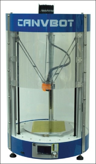
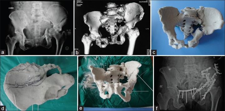
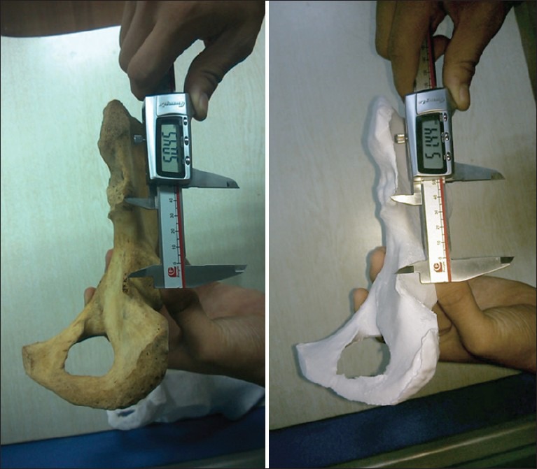
First, 16 dried human cadaveric pelvises were used to confirm the anatomical accuracy of the 3D models printed based on radiographic data. Next, nine clinical cases between January 2009 and April 2013 were used to evaluate the surgical reconstruction based on the 3D printed models. The pelvic injuries were all type C, and the average time from injury to reconstruction was 11 weeks (range: 8-17 weeks). The workflow consisted of: (1) Printing patient-specific bone models based on preoperative computed tomography (CT) scans, (2) virtual fracture reduction using the printed 3D anatomic template, (3) virtual fracture fixation using Kirschner wires, and (4) preoperatively measuring the osteotomy and implant position relative to landmarks using the virtually defined deformation. These models aided communication between surgical team members during the procedure. This technique was validated by comparing the preoperative planning to the intraoperative procedure.
The accuracy of the 3D printed models was within specification. Production of a model from standard CT DICOM data took 7 hours (range: 6-9 hours). Preoperative planning using the 3D printed models was feasible in all cases. Good correlation was found between the preoperative planning and postoperative follow-up X-ray in all nine cases. The patients were followed for 3-29 months (median: 5 months). The fracture healing time was 9-17 weeks (mean: 10 weeks). No delayed incision healing, wound infection, or nonunions occurred. The results were excellent in two cases, good in five, and poor in two based on the Majeed score.
The 3D printing planning technique for pelvic surgery was successfully integrated into a clinical workflow to improve patient-specific preoperative planning by providing a visual and haptic model of the injury and allowing patient-specific adaptation of each osteosynthesis implant to the virtually reduced pelvis.
陈旧性骨盆骨折是最难治疗的骨折之一,因其解剖结构复杂、手术部位难以触及且此类病例发病率相对较低。要实现骨盆环对称及骨折的稳定固定,进行恰当的评估和手术规划很有必要。本研究的目的是评估三维(3D)打印技术在陈旧性骨盆骨折手术治疗中的应用。
首先,使用16具干燥的人体尸体骨盆来确认基于放射学数据打印的3D模型的解剖准确性。接下来,选取2009年1月至2013年4月间的9例临床病例,基于3D打印模型评估手术重建情况。骨盆损伤均为C型,受伤至重建的平均时间为11周(范围:8 - 17周)。工作流程包括:(1)根据术前计算机断层扫描(CT)扫描打印患者特异性骨模型;(2)使用打印的3D解剖模板进行虚拟骨折复位;(3)使用克氏针进行虚拟骨折固定;(4)术前使用虚拟定义的变形测量截骨术及植入物相对于标志点的位置。这些模型有助于手术团队成员在手术过程中的沟通。通过将术前规划与术中操作进行比较来验证该技术。
3D打印模型的准确性符合规格。从标准CT DICOM数据生成一个模型耗时7小时(范围:6 - 9小时)。在所有病例中,使用3D打印模型进行术前规划都是可行的。在所有9例病例中,术前规划与术后随访X线片之间均发现良好的相关性。对患者进行了3 - 29个月的随访(中位数:5个月)。骨折愈合时间为9 - 17周(平均:10周)。未发生切口延迟愈合、伤口感染或骨不连。根据马吉德评分,2例结果为优,5例为良,2例为差。
骨盆手术的3D打印规划技术成功整合到临床工作流程中,通过提供损伤的视觉和触觉模型,并允许根据患者特异性对每个接骨植入物进行调整以适应虚拟复位的骨盆,从而改善了患者特异性术前规划。