Ji Guangchen, Li Zhen, Neugebauer Volker
Department of Pharmacology and Neuroscience, School of Medicine, Texas Tech University Health Sciences Center (TTUHSC), Lubbock, TX Institute for Biomedical Sciences of Pain and Institute for Functional Brain Disorders, The Fourth Military Medical University, Xi'an, China.
Pain. 2015 May;156(5):825-836. doi: 10.1097/j.pain.0000000000000120.
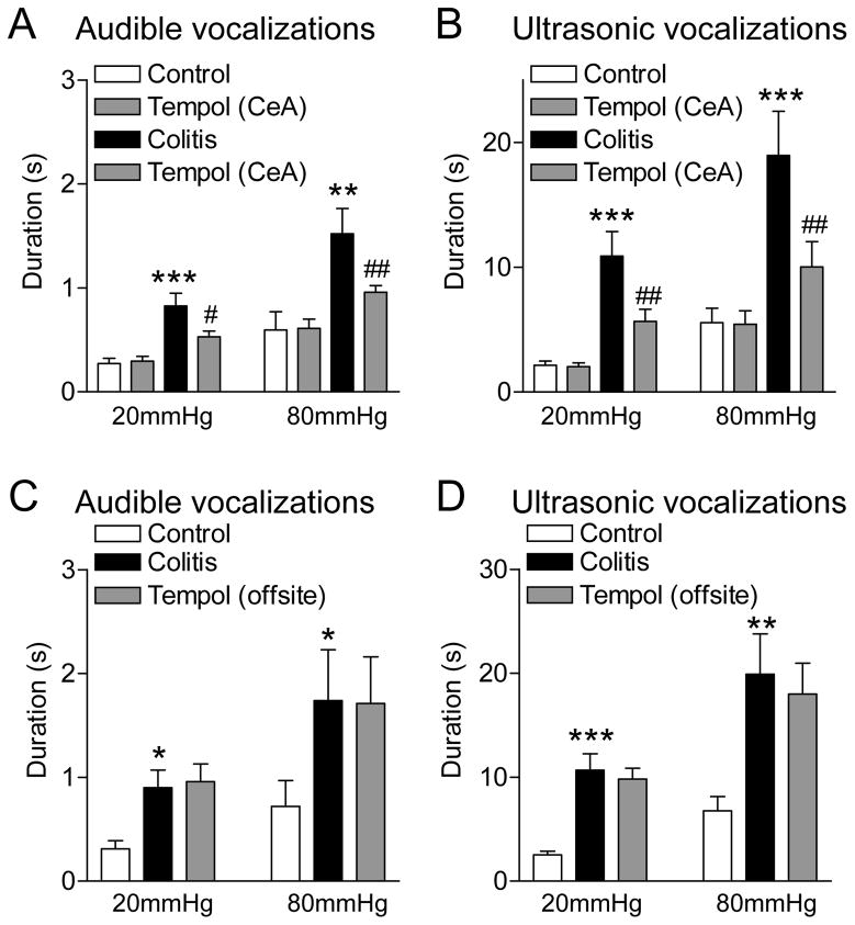
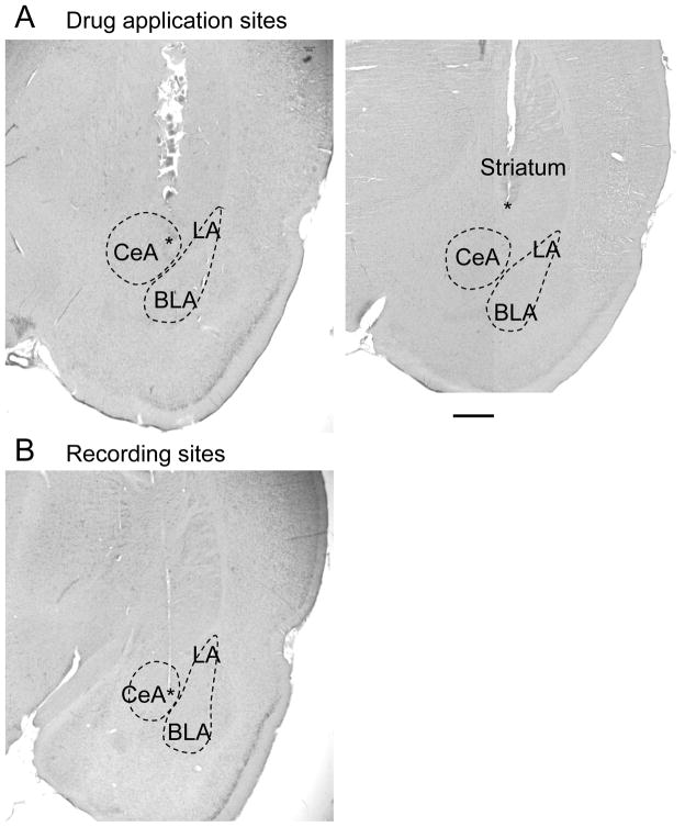
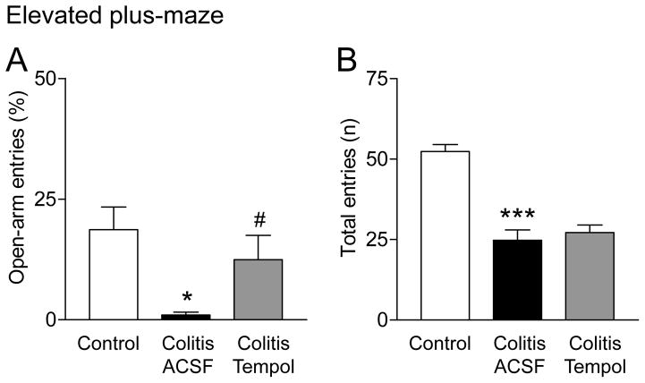
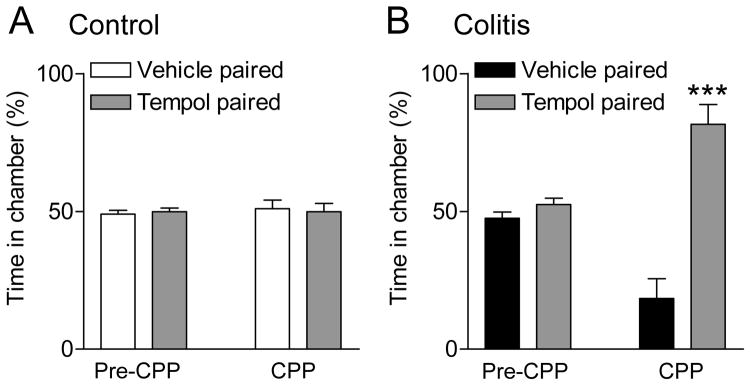
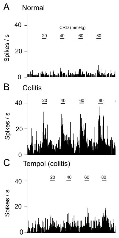
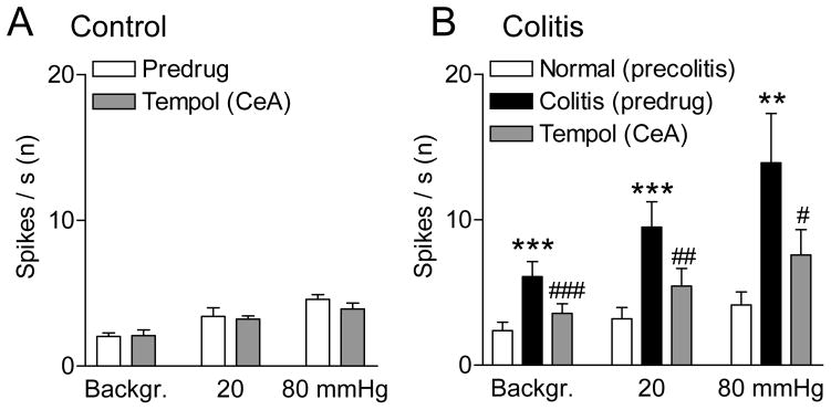
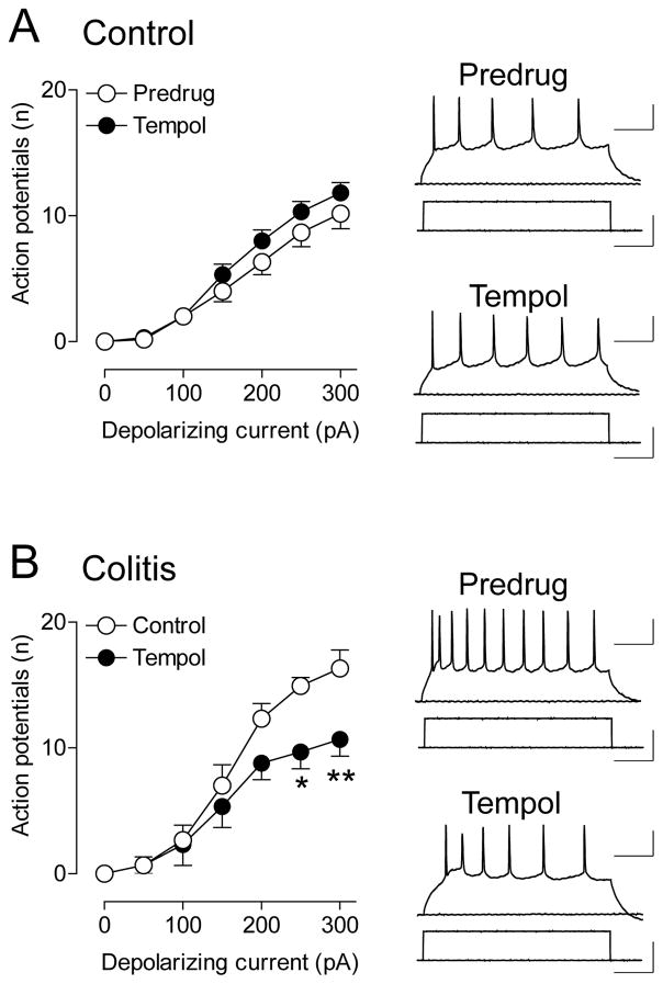
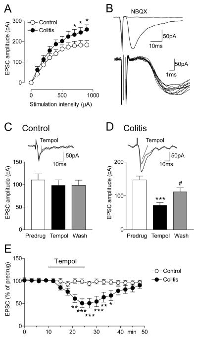
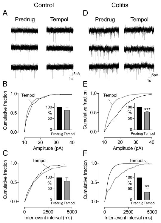
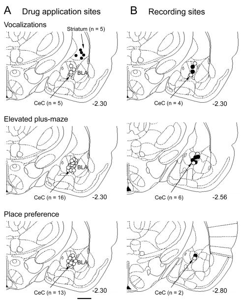
Accumulating evidence suggests an important contribution of reactive oxygen species (ROS) to pain and neuropsychiatric disorders, but their role in pain-related plasticity in the brain is largely unknown. Neuroplasticity in the central nucleus of the amygdala (CeA) correlates positively with pain behaviors in different models. Little is known, however, about mechanisms of visceral pain-related amygdala changes. The electrophysiological and behavioral studies reported here addressed the role of ROS in the CeA in a visceral pain model induced by intracolonic zymosan. Vocalizations to colorectal distension and anxiety-like behavior increased after intracolonic zymosan and were inhibited by intra-CeA application of a ROS scavenger (tempol, a superoxide dismutase mimetic). Tempol also induced a place preference in zymosan-treated rats but not in controls. Single-unit recordings of CeA neurons in anesthetized rats showed increases of background activity and responses to visceral stimuli after intracolonic zymosan. Intra-CeA application of tempol inhibited the increased activity but had no effect under normal conditions. Whole-cell patch-clamp recordings of CeA neurons in brain slices from zymosan-treated rats showed that tempol decreased neuronal excitability and excitatory synaptic transmission of presumed nociceptive inputs from the brainstem (parabrachial area) through a combination of presynaptic and postsynaptic actions. Tempol had no effect in brain slices from sham controls. The results suggest that ROS contribute to visceral pain-related hyperactivity of amygdala neurons and amygdala-dependent behaviors through a mechanism that involves increased excitatory transmission and excitability of CeA neurons.
越来越多的证据表明,活性氧(ROS)在疼痛和神经精神疾病中起着重要作用,但其在大脑疼痛相关可塑性中的作用仍 largely unknown。杏仁核中央核(CeA)的神经可塑性在不同模型中与疼痛行为呈正相关。然而,关于内脏痛相关杏仁核变化的机制却知之甚少。本文报道的电生理和行为学研究探讨了ROS在结肠内注射酵母聚糖诱导的内脏痛模型中CeA的作用。结肠内注射酵母聚糖后,对结直肠扩张的发声和焦虑样行为增加,并被CeA内注射ROS清除剂(tempol,一种超氧化物歧化酶模拟物)所抑制。Tempol还在酵母聚糖处理的大鼠中诱导了位置偏好,但在对照组中没有。对麻醉大鼠CeA神经元的单单位记录显示,结肠内注射酵母聚糖后,背景活动和对内脏刺激的反应增加。CeA内注射tempol可抑制这种增加的活动,但在正常条件下没有影响。对酵母聚糖处理大鼠脑片上CeA神经元的全细胞膜片钳记录显示,tempol通过突触前和突触后作用的组合,降低了假定来自脑干(臂旁区)的伤害性输入的神经元兴奋性和兴奋性突触传递。Tempol对假手术对照组的脑片没有影响。结果表明,ROS通过一种涉及CeA神经元兴奋性传递和兴奋性增加的机制,导致杏仁核神经元与内脏痛相关的活动亢进和杏仁核依赖性行为。