Division of Mood Disorders, Psychiatry Department, Shanghai Mental Health Center, Shanghai Jiaotong University, Shanghai, China.
Key Laboratory of Animal Models and Human Disease Mechanisms of the Chinese Academy of Sciences and Yunnan Province, Kunming Institute of Zoology, Kunming, China.
Psychiatry Investig. 2015 Apr;12(2):227-34. doi: 10.4306/pi.2015.12.2.227. Epub 2015 Feb 2.
Evidence of the brain network involved in cognitive dysfunction has been inconsistent for major depressive disorder (MDD), especially during early stage of MDD. This study seeks to examine abnormal cognition connectivity network (CCN) in MDD within the whole brain.
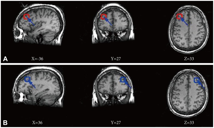
Sixteen patients with MDD and 16 health controls were scanned during resting-state using 3.0 T functional magnetic resonance imaging (fMRI). All patients were first episode without any history of antidepressant treatment. Both the left and right dorsolateral prefrontal cortex (DLPFC) were used as individual seeds to identify CCN by the seed-target correlation analysis. Two sample t test was used to calculate between-group differences in CCN using fisher z-transformed correlation maps.
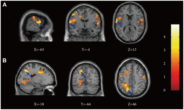
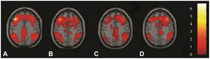
The CCN was constructed by bilateral seed DLPFC in two groups separately. Depressed subjects exhibited significantly increased functional connectivity (FC) by left DLPFC in one cluster, overlapping middle frontal gyrus, BA7, BA43, precuneus, BA6, BA40, superior temporal gyrus, BA22, inferior parietal lobule, precentral gyrus, BA4 and cingulate gyrus in left cerebrum. Health controls did not show any cluster with significantly greater FC compared to depressed subjects in left DLPFC network. There was no significant difference of FC in right DLPFC network between depressed subjects and the health controls.
There are differences in CCN during early stage of MDD, as identified by increased FCs among part of frontal gyrus, parietal cortex, cingulate cortex, and BA43, BA22, BA4 with left DLPFC. These brain areas might be involved in the underlying mechanisms of cognitive dysfunction in MDD.
有证据表明,重度抑郁症(MDD)患者的认知功能障碍的脑网络一直存在不一致的情况,尤其是在 MDD 的早期阶段。本研究旨在探讨 MDD 患者全脑范围内的异常认知连接网络(CCN)。
16 名 MDD 患者和 16 名健康对照者在静息状态下使用 3.0T 功能磁共振成像(fMRI)进行扫描。所有患者均为首次发作,且均未接受过抗抑郁治疗。使用左、右侧背外侧前额叶皮层(DLPFC)作为个体种子,通过种子-目标相关分析来识别 CCN。采用两样本 t 检验,对经 fisher z 转换的相关图进行组间差异分析。
两组分别通过双侧种子 DLPFC 构建 CCN。与健康对照组相比,抑郁组患者左 DLPFC 一个脑区的功能连接(FC)显著增加,重叠区域包括额中回、额下回、扣带回、顶下小叶、颞上回、角回、顶下小叶、中央前回和扣带回。健康对照组的左 DLPFC 网络中没有任何脑区显示出与抑郁组相比具有更高 FC 的显著差异。抑郁组和健康对照组右 DLPFC 网络的 FC 无显著差异。
在 MDD 的早期阶段,存在 CCN 的差异,左 DLPFC 与部分额回、顶叶皮层、扣带回以及 BA43、BA22、BA4 的 FC 增加有关。这些脑区可能与 MDD 认知功能障碍的潜在机制有关。