Han Myounghee, Kim Kyunghun, Jang Sun-Joo, Cho Han Saem, Bouma Brett E, Oh Wang-Yuhl, Ryu Sukyoung
Department of Computer Science, Korea Advanced Institute of Science and Technology, Daejeon, Republic of Korea.
Department of Mechanical Engineering, Korea Advanced Institute of Science and Technology, Daejeon, Republic of Korea; Graduate School of Medical Science and Engineering, Korea Advanced Institute of Science and Technology, Daejeon, Republic of Korea.
PLoS One. 2015 Apr 16;10(4):e0124192. doi: 10.1371/journal.pone.0124192. eCollection 2015.
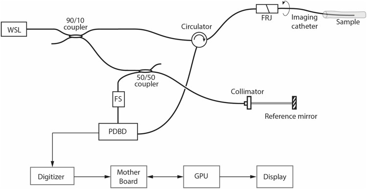
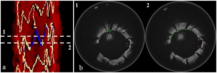
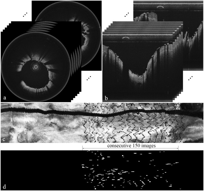
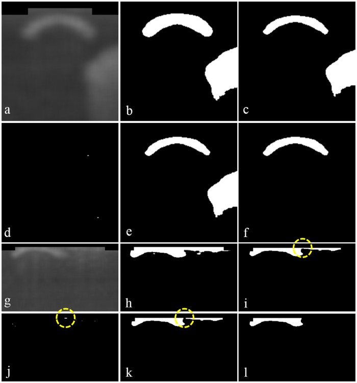
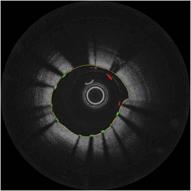
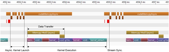
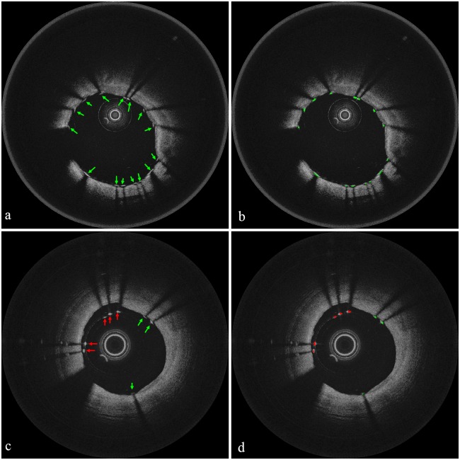
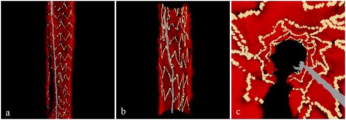
Frequency domain optical coherence tomography (FD-OCT) has become one of the important clinical tools for intracoronary imaging to diagnose and monitor coronary artery disease, which has been one of the leading causes of death. To help more accurate diagnosis and monitoring of the disease, many researchers have recently worked on visualization of various coronary microscopic features including stent struts by constructing three-dimensional (3D) volumetric rendering from series of cross-sectional intracoronary FD-OCT images. In this paper, we present the first, to our knowledge, "push-of-a-button" graphics processing unit (GPU)-accelerated framework for intracoronary OCT imaging. Our framework visualizes 3D microstructures of the vessel wall with stent struts from raw binary OCT data acquired by the system digitizer as one seamless process. The framework reports the state-of-the-art performance; from raw OCT data, it takes 4.7 seconds to provide 3D visualization of a 5-cm-long coronary artery (of size 1600 samples x 1024 A-lines x 260 frames) with stent struts and detection of malapposition automatically at the single push of a button.
频域光学相干断层扫描(FD-OCT)已成为冠状动脉内成像诊断和监测冠状动脉疾病的重要临床工具之一,冠状动脉疾病一直是主要死因之一。为了更准确地诊断和监测该疾病,最近许多研究人员致力于通过从一系列冠状动脉内FD-OCT横截面图像构建三维(3D)体积渲染来可视化包括支架支柱在内的各种冠状动脉微观特征。在本文中,据我们所知,我们提出了首个用于冠状动脉内OCT成像的“一键式”图形处理单元(GPU)加速框架。我们的框架将系统数字化仪采集的原始二进制OCT数据中的血管壁3D微观结构与支架支柱作为一个无缝过程进行可视化。该框架展现了最先进的性能;从原始OCT数据开始,只需一键操作,就能在4.7秒内提供一条5厘米长的带有支架支柱的冠状动脉(尺寸为1600个样本×1024条A线×260帧)的3D可视化图像,并自动检测贴壁不良情况。