• 文献检索
  • 文档翻译
  • 深度研究
  • 学术资讯
  • Suppr Zotero 插件Zotero 插件
  • 邀请有礼
  • 套餐&价格
  • 历史记录
应用&插件
Suppr Zotero 插件Zotero 插件浏览器插件Mac 客户端Windows 客户端微信小程序
定价
高级版会员购买积分包购买API积分包
服务
文献检索文档翻译深度研究API 文档MCP 服务
关于我们
关于 Suppr公司介绍联系我们用户协议隐私条款
关注我们

Suppr 超能文献

核心技术专利:CN118964589B侵权必究
粤ICP备2023148730 号-1Suppr @ 2026

文献检索

告别复杂PubMed语法,用中文像聊天一样搜索,搜遍4000万医学文献。AI智能推荐,让科研检索更轻松。

立即免费搜索

文件翻译

保留排版,准确专业,支持PDF/Word/PPT等文件格式,支持 12+语言互译。

免费翻译文档

深度研究

AI帮你快速写综述,25分钟生成高质量综述,智能提取关键信息,辅助科研写作。

立即免费体验

肌动蛋白样ParM细丝的结构显示了质粒分离纺锤体的结构。

Structures of actin-like ParM filaments show architecture of plasmid-segregating spindles.

作者信息

Bharat Tanmay A M, Murshudov Garib N, Sachse Carsten, Löwe Jan

机构信息

Structural Studies Division, MRC Laboratory of Molecular Biology, Francis Crick Avenue, Cambridge CB2 0QH, UK.

Structural and Computational Biology Unit, European Molecular Biology Laboratory, Meyerhofstrasse 1, Heidelberg 69117, Germany.

出版信息

Nature. 2015 Jul 2;523(7558):106-10. doi: 10.1038/nature14356. Epub 2015 Apr 27.

DOI:10.1038/nature14356
PMID:25915019
原文链接:https://pmc.ncbi.nlm.nih.gov/articles/PMC4493928/
Abstract

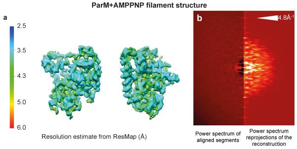
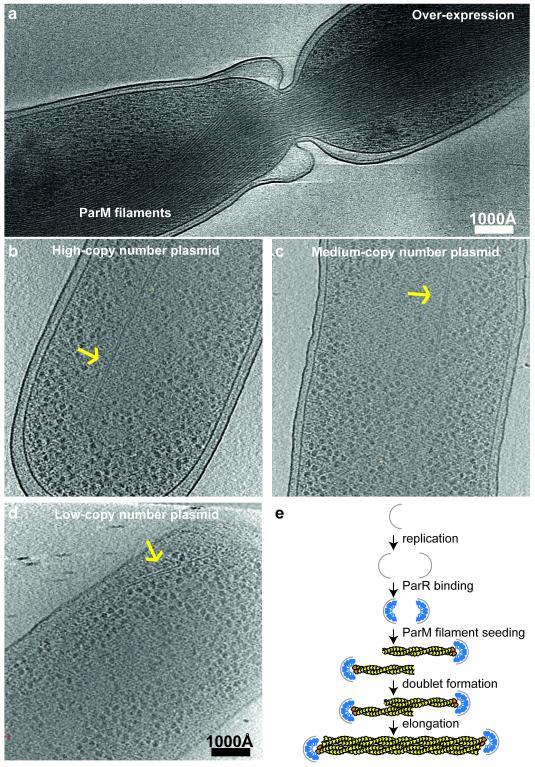
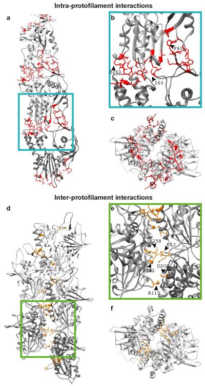
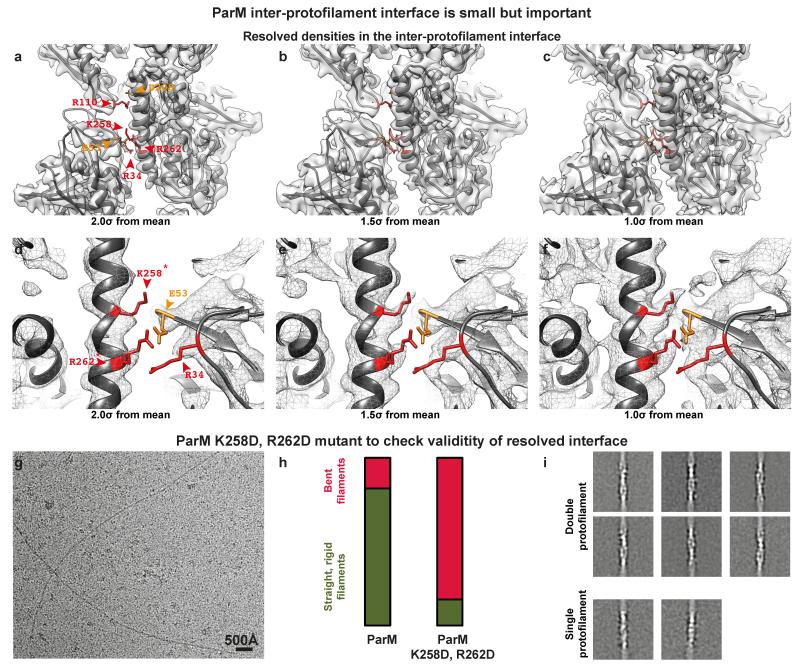
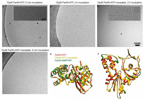
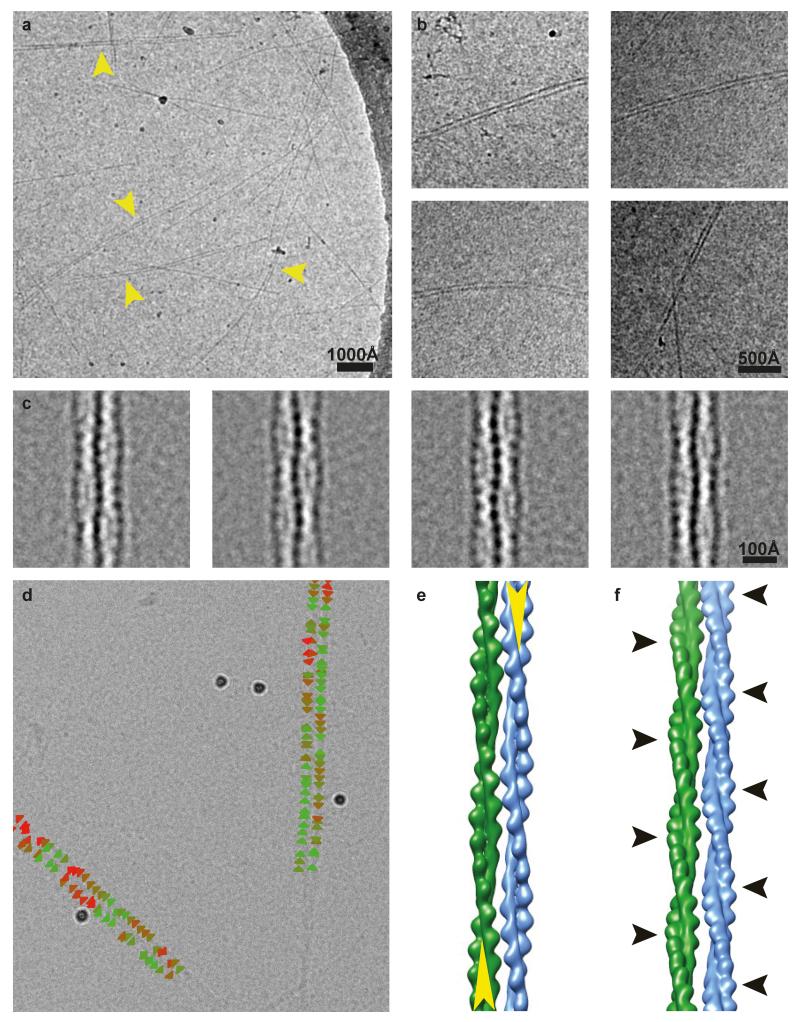
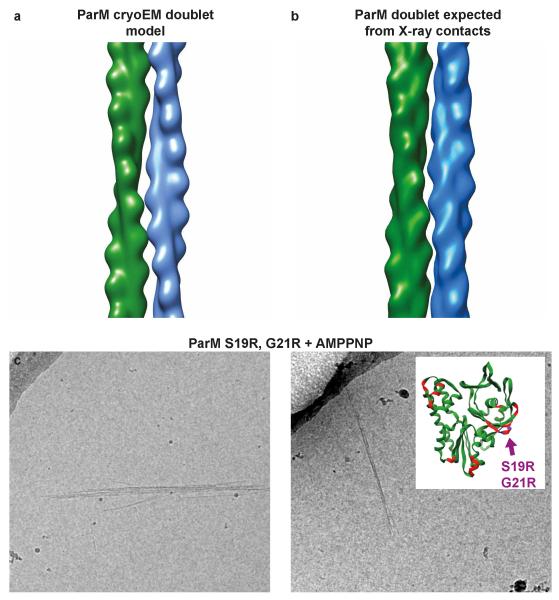
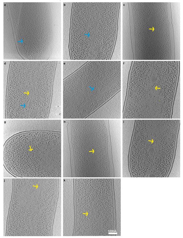
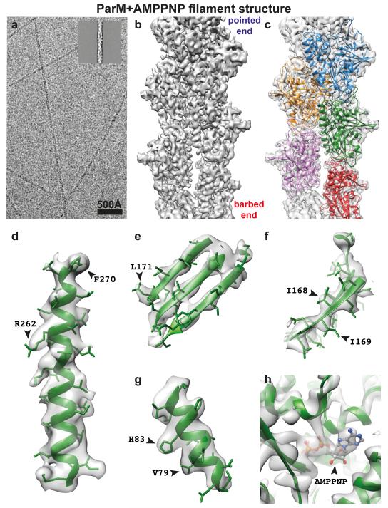
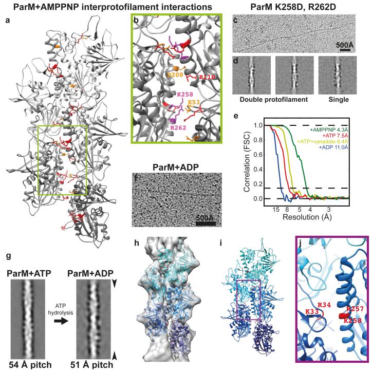
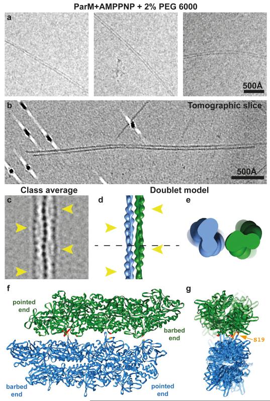
Active segregation of Escherichia coli low-copy-number plasmid R1 involves formation of a bipolar spindle made of left-handed double-helical actin-like ParM filaments. ParR links the filaments with centromeric parC plasmid DNA, while facilitating the addition of subunits to ParM filaments. Growing ParMRC spindles push sister plasmids to the cell poles. Here, using modern electron cryomicroscopy methods, we investigate the structures and arrangements of ParM filaments in vitro and in cells, revealing at near-atomic resolution how subunits and filaments come together to produce the simplest known mitotic machinery. To understand the mechanism of dynamic instability, we determine structures of ParM filaments in different nucleotide states. The structure of filaments bound to the ATP analogue AMPPNP is determined at 4.3 Å resolution and refined. The ParM filament structure shows strong longitudinal interfaces and weaker lateral interactions. Also using electron cryomicroscopy, we reconstruct ParM doublets forming antiparallel spindles. Finally, with whole-cell electron cryotomography, we show that doublets are abundant in bacterial cells containing low-copy-number plasmids with the ParMRC locus, leading to an asynchronous model of R1 plasmid segregation.

摘要

大肠杆菌低拷贝数质粒R1的主动分离涉及由左手双螺旋肌动蛋白样ParM细丝构成的双极纺锤体的形成。ParR将细丝与着丝粒parC质粒DNA相连,同时促进亚基添加到ParM细丝上。生长中的ParMRC纺锤体将姐妹质粒推向细胞两极。在此,我们使用现代电子冷冻显微镜方法研究了体外和细胞内ParM细丝的结构与排列,以近原子分辨率揭示了亚基和细丝如何聚集在一起形成已知最简单的有丝分裂机制。为了解动态不稳定性的机制,我们确定了处于不同核苷酸状态的ParM细丝的结构。与ATP类似物AMPPNP结合的细丝结构在4.3 Å分辨率下确定并得到优化。ParM细丝结构显示出强纵向界面和较弱的横向相互作用。我们还利用电子冷冻显微镜重建了形成反平行纺锤体的ParM双联体。最后,通过全细胞电子冷冻断层扫描,我们表明双联体在含有具有ParMRC位点的低拷贝数质粒的细菌细胞中大量存在,从而得出R1质粒分离的异步模型。

https://cdn.ncbi.nlm.nih.gov/pmc/blobs/c80f/4493928/fc006ae2e682/emss-62410-f0011.jpg
https://cdn.ncbi.nlm.nih.gov/pmc/blobs/c80f/4493928/546e00c5a279/emss-62410-f0001.jpg
https://cdn.ncbi.nlm.nih.gov/pmc/blobs/c80f/4493928/bb876f314eed/emss-62410-f0002.jpg
https://cdn.ncbi.nlm.nih.gov/pmc/blobs/c80f/4493928/a98c1c734ff5/emss-62410-f0003.jpg
https://cdn.ncbi.nlm.nih.gov/pmc/blobs/c80f/4493928/9cba02eeb1fa/emss-62410-f0004.jpg
https://cdn.ncbi.nlm.nih.gov/pmc/blobs/c80f/4493928/13ee2aaa16ce/emss-62410-f0005.jpg
https://cdn.ncbi.nlm.nih.gov/pmc/blobs/c80f/4493928/72974b9fdcbb/emss-62410-f0006.jpg
https://cdn.ncbi.nlm.nih.gov/pmc/blobs/c80f/4493928/2c71cfd53534/emss-62410-f0007.jpg
https://cdn.ncbi.nlm.nih.gov/pmc/blobs/c80f/4493928/a9d5c4744ae4/emss-62410-f0008.jpg
https://cdn.ncbi.nlm.nih.gov/pmc/blobs/c80f/4493928/111dc488fab9/emss-62410-f0009.jpg
https://cdn.ncbi.nlm.nih.gov/pmc/blobs/c80f/4493928/60db7598a8d6/emss-62410-f0010.jpg
https://cdn.ncbi.nlm.nih.gov/pmc/blobs/c80f/4493928/fc006ae2e682/emss-62410-f0011.jpg

相似文献

1
Structures of actin-like ParM filaments show architecture of plasmid-segregating spindles.肌动蛋白样ParM细丝的结构显示了质粒分离纺锤体的结构。
Nature. 2015 Jul 2;523(7558):106-10. doi: 10.1038/nature14356. Epub 2015 Apr 27.
2
A bipolar spindle of antiparallel ParM filaments drives bacterial plasmid segregation.双极纺锤体的反平行 ParM 丝驱动细菌质粒分离。
Science. 2012 Dec 7;338(6112):1334-7. doi: 10.1126/science.1229091. Epub 2012 Oct 25.
3
Electron cryomicroscopy of E. coli reveals filament bundles involved in plasmid DNA segregation.大肠杆菌的电子冷冻显微镜显示参与质粒DNA分离的丝状束。
Science. 2009 Jan 23;323(5913):509-12. doi: 10.1126/science.1164346. Epub 2008 Dec 18.
4
Bacterial actin: architecture of the ParMRC plasmid DNA partitioning complex.细菌肌动蛋白:ParMRC 质粒 DNA 分配复合物的结构
EMBO J. 2008 Aug 20;27(16):2230-8. doi: 10.1038/emboj.2008.152. Epub 2008 Jul 24.
5
F-actin-like filaments formed by plasmid segregation protein ParM.由质粒分离蛋白ParM形成的类F-肌动蛋白丝。
EMBO J. 2002 Dec 16;21(24):6935-43. doi: 10.1093/emboj/cdf672.
6
Cryo-EM reconstruction of AlfA from reveals the structure of a simplified actin-like filament at 3.4-Å resolution.Cryo-EM 重建揭示了 AlfA 在 3.4-Å 分辨率下的简化肌动蛋白样丝结构。
Proc Natl Acad Sci U S A. 2018 Mar 27;115(13):3458-3463. doi: 10.1073/pnas.1716424115. Epub 2018 Feb 13.
7
The structure of bacterial ParM filaments.细菌ParM细丝的结构。
Nat Struct Mol Biol. 2007 Oct;14(10):921-6. doi: 10.1038/nsmb1300. Epub 2007 Sep 16.
8
The ParMRC system: molecular mechanisms of plasmid segregation by actin-like filaments.ParMRC 系统:肌动蛋白样丝分裂质粒分离的分子机制。
Nat Rev Microbiol. 2010 Oct;8(10):683-92. doi: 10.1038/nrmicro2425.
9
Molecular structure of the ParM polymer and the mechanism leading to its nucleotide-driven dynamic instability.ParM聚合物的分子结构及其核苷酸驱动的动态不稳定性的机制。
EMBO J. 2008 Feb 6;27(3):570-9. doi: 10.1038/sj.emboj.7601978. Epub 2008 Jan 10.
10
Protein-nanocrystal conjugates support a single filament polymerization model in R1 plasmid segregation.蛋白质-纳米晶体共轭物支持R1质粒分离中的单丝聚合模型。
J Biol Chem. 2008 Oct 17;283(42):28081-6. doi: 10.1074/jbc.M803833200. Epub 2008 Jul 24.

引用本文的文献

1
Bacterial genome-encoded ParMs.细菌基因组编码的ParM蛋白。
J Biol Chem. 2025 Jul;301(7):110351. doi: 10.1016/j.jbc.2025.110351. Epub 2025 Jun 9.
2
Actin network evolution as a key driver of eukaryotic diversification.肌动蛋白网络进化作为真核生物多样化的关键驱动力。
J Cell Sci. 2024 Aug 1;137(15). doi: 10.1242/jcs.261660. Epub 2024 Aug 9.
3
DNA Segregation in Enterobacteria.肠杆菌中的DNA分离

本文引用的文献

1
Near-atomic resolution for one state of F-actin.丝状肌动蛋白一种状态的近原子分辨率。
Structure. 2015 Jan 6;23(1):173-182. doi: 10.1016/j.str.2014.11.006. Epub 2014 Dec 18.
2
Structure of the F-actin-tropomyosin complex.F-肌动蛋白-原肌球蛋白复合物的结构。
Nature. 2015 Mar 5;519(7541):114-7. doi: 10.1038/nature14033. Epub 2014 Dec 1.
3
High-resolution microtubule structures reveal the structural transitions in αβ-tubulin upon GTP hydrolysis.高分辨率微管结构揭示了 GTP 水解时 αβ-微管蛋白的结构转变。
EcoSal Plus. 2023 Dec 12;11(1):eesp00382020. doi: 10.1128/ecosalplus.esp-0038-2020. Epub 2023 May 9.
4
Single particle cryo-EM analysis of Rickettsia conorii Sca2 reveals a formin-like core.单细胞冷冻电镜分析表明恙虫病东方体 Sca2 存在类似formin 的核心结构。
J Struct Biol. 2023 Jun;215(2):107960. doi: 10.1016/j.jsb.2023.107960. Epub 2023 Apr 5.
5
Diverse cytomotive actins and tubulins share a polymerization switch mechanism conferring robust dynamics.不同的细胞运动肌动蛋白和微管蛋白共享一个聚合开关机制,赋予其强大的动力学特性。
Sci Adv. 2023 Mar 29;9(13):eadf3021. doi: 10.1126/sciadv.adf3021.
6
Structural mechanism for bidirectional actin cross-linking by T-plastin.T 型蛋白通过形成二聚体结构实现对肌动蛋白的双向交联
Proc Natl Acad Sci U S A. 2022 Sep 13;119(37):e2205370119. doi: 10.1073/pnas.2205370119. Epub 2022 Sep 6.
7
The Specificity of ParR Binding Determines the Incompatibility of Conjugative Plasmids in Clostridium perfringens.ParR 结合的特异性决定了产气荚膜梭菌中可移动质粒的不相容性。
mBio. 2022 Aug 30;13(4):e0135622. doi: 10.1128/mbio.01356-22. Epub 2022 Jun 21.
8
: a neural-network-based approach for identification of unknown proteins in X-ray crystallography and cryo-EM.一种基于神经网络的方法,用于在X射线晶体学和冷冻电镜中鉴定未知蛋白质。
IUCrJ. 2021 Dec 1;9(Pt 1):86-97. doi: 10.1107/S2052252521011088. eCollection 2022 Jan 1.
9
The C-terminal region of the plasmid partitioning protein TubY is a tetramer that can bind membranes and DNA.质粒分配蛋白 TubY 的 C 端区域是一个可以结合膜和 DNA 的四聚体。
J Biol Chem. 2020 Dec 18;295(51):17770-17780. doi: 10.1074/jbc.RA120.014705.
10
Unique Properties of the Alpha-Helical DNA-Binding Protein KfrA Encoded by the IncU Incompatibility Group Plasmid RA3 and Its Host-Dependent Role in Plasmid Maintenance.由 IncU 不相容群质粒 RA3 编码的α-螺旋 DNA 结合蛋白 KfrA 的独特性质及其在质粒维持中的宿主依赖性作用。
Appl Environ Microbiol. 2021 Jan 4;87(2). doi: 10.1128/AEM.01771-20.
Cell. 2014 May 22;157(5):1117-29. doi: 10.1016/j.cell.2014.03.053.
4
Crenactin from Pyrobaculum calidifontis is closely related to actin in structure and forms steep helical filaments.来自喜热裂古菌的 Crenactin 在结构上与肌动蛋白密切相关,并形成陡峭的螺旋丝。
FEBS Lett. 2014 Mar 3;588(5):776-82. doi: 10.1016/j.febslet.2014.01.029. Epub 2014 Jan 28.
5
Bacterial actins and their diversity.细菌肌动蛋白及其多样性。
Biochemistry. 2013 Oct 8;52(40):6928-39. doi: 10.1021/bi4010792. Epub 2013 Sep 24.
6
The bacterial actin MamK: in vitro assembly behavior and filament architecture.细菌肌动蛋白 MamK:体外组装行为和丝状体结构。
J Biol Chem. 2013 Feb 8;288(6):4265-77. doi: 10.1074/jbc.M112.417030. Epub 2012 Nov 30.
7
A bipolar spindle of antiparallel ParM filaments drives bacterial plasmid segregation.双极纺锤体的反平行 ParM 丝驱动细菌质粒分离。
Science. 2012 Dec 7;338(6112):1334-7. doi: 10.1126/science.1229091. Epub 2012 Oct 25.
8
Direct visualization of secondary structures of F-actin by electron cryomicroscopy.电子冷冻显微镜直接观察 F-肌动蛋白的二级结构。
Nature. 2010 Oct 7;467(7316):724-8. doi: 10.1038/nature09372. Epub 2010 Sep 15.
9
Pushing and pulling in prokaryotic DNA segregation.在原核生物 DNA 分离中推和拉。
Cell. 2010 Jun 11;141(6):927-42. doi: 10.1016/j.cell.2010.05.033.
10
Molecular mechanism of bundle formation by the bacterial actin ParM.细菌肌动蛋白 ParM 形成束的分子机制。
Biochem Biophys Res Commun. 2010 Jan 22;391(4):1598-603. doi: 10.1016/j.bbrc.2009.12.078. Epub 2009 Dec 22.