Lou Sunny S, Diz-Muñoz Alba, Weiner Orion D, Fletcher Daniel A, Theriot Julie A
Department of Chemical and Systems Biology, Department of Biochemistry, and Howard Hughes Medical Institute, Stanford University School of Medicine, Stanford, CA 94305.
Department of Bioengineering and Biophysics Program, University of California, Berkeley, Berkeley, CA 94720 Department of Bioengineering and Biophysics Program, University of California, Berkeley, Berkeley, CA 94720 Cardiovascular Research Institute and Department of Biochemistry, University of California, San Francisco, San Francisco, CA 94158 Cardiovascular Research Institute and Department of Biochemistry, University of California, San Francisco, San Francisco, CA 94158.
J Cell Biol. 2015 Apr 27;209(2):275-88. doi: 10.1083/jcb.201409001.
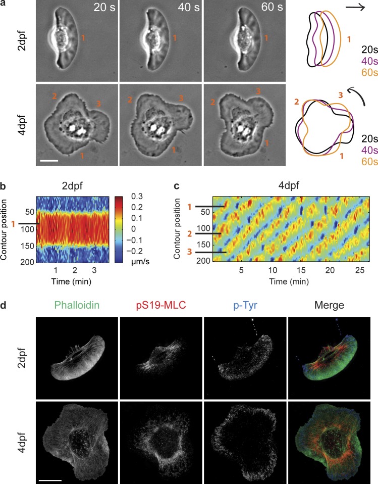
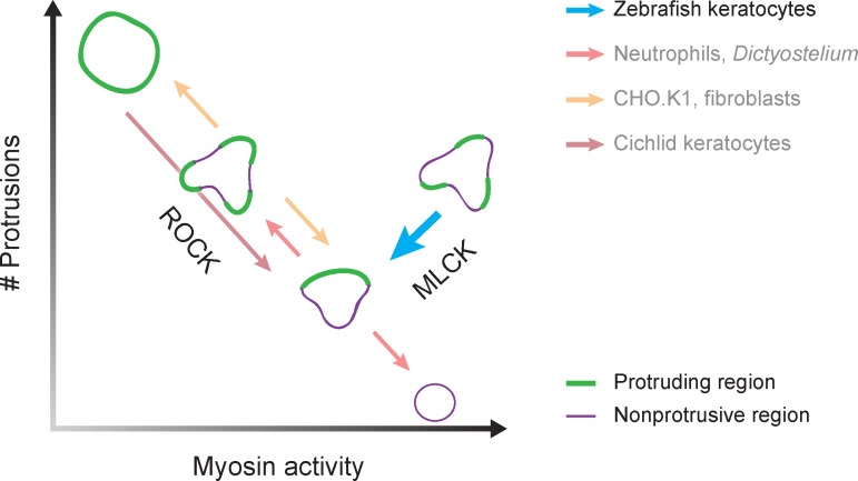
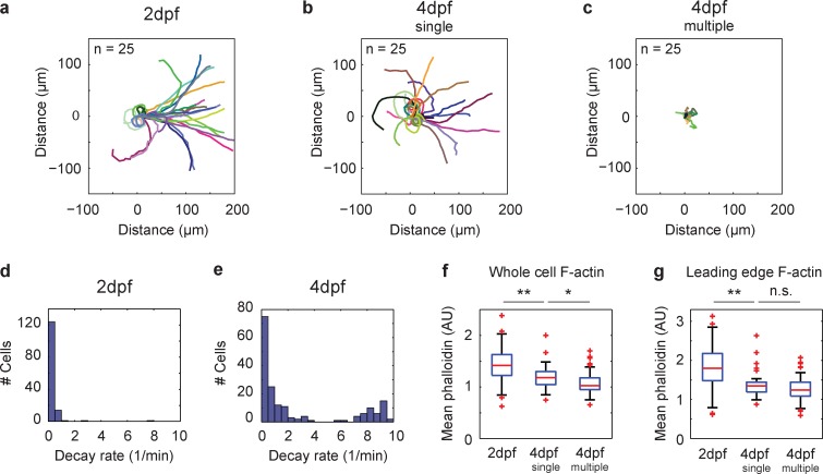
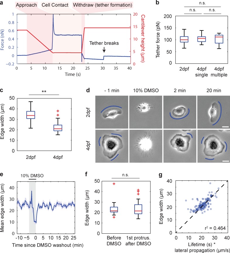
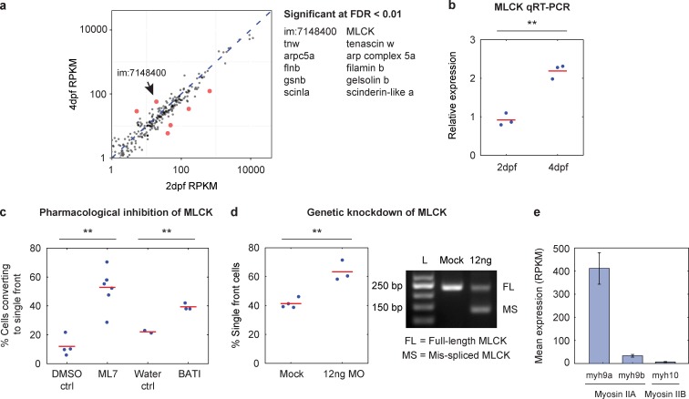
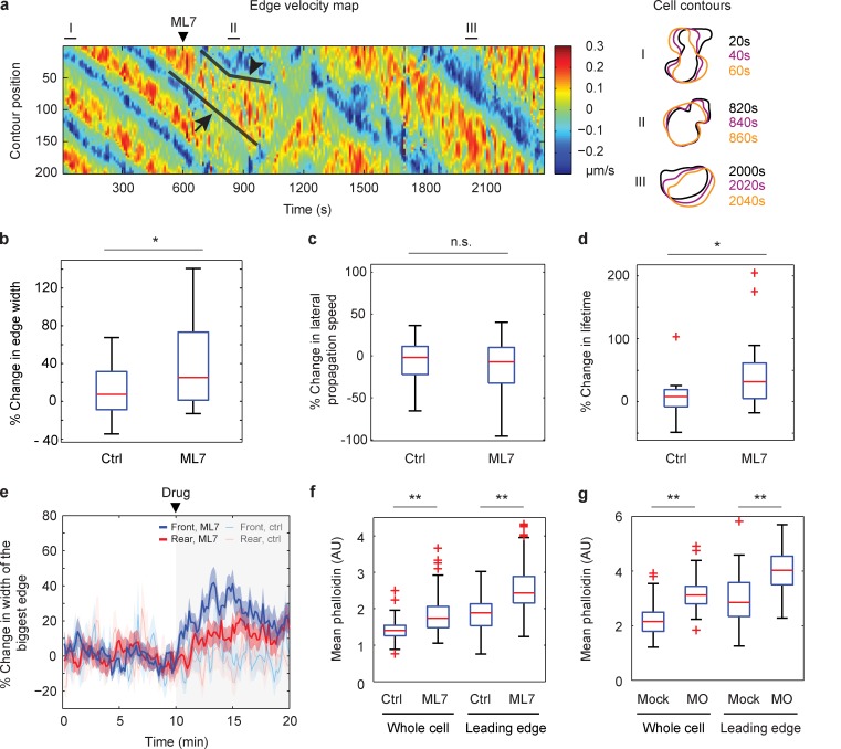
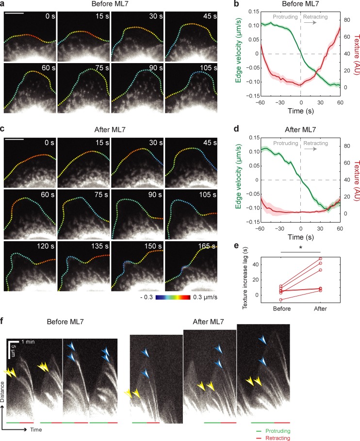
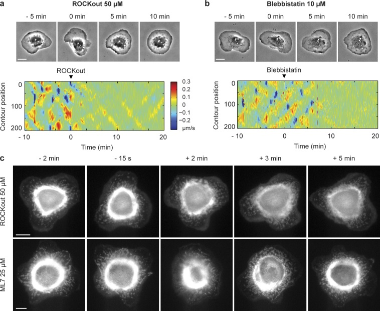
Cells polarize to a single front and rear to achieve rapid actin-based motility, but the mechanisms preventing the formation of multiple fronts are unclear. We developed embryonic zebrafish keratocytes as a model system for investigating establishment of a single axis. We observed that, although keratocytes from 2 d postfertilization (dpf) embryos resembled canonical fan-shaped keratocytes, keratocytes from 4 dpf embryos often formed multiple protrusions despite unchanged membrane tension. Using genomic, genetic, and pharmacological approaches, we determined that the multiple-protrusion phenotype was primarily due to increased myosin light chain kinase (MLCK) expression. MLCK activity influences cell polarity by increasing myosin accumulation in lamellipodia, which locally decreases protrusion lifetime, limiting lamellipodial size and allowing for multiple protrusions to coexist within the context of membrane tension limiting protrusion globally. In contrast, Rho kinase (ROCK) regulates myosin accumulation at the cell rear and does not determine protrusion size. These results suggest a novel MLCK-specific mechanism for controlling cell polarity via regulation of myosin activity in protrusions.
细胞极化形成单一的前端和后端以实现基于肌动蛋白的快速运动,但阻止形成多个前端的机制尚不清楚。我们开发了胚胎斑马鱼角质形成细胞作为研究单轴建立的模型系统。我们观察到,虽然受精后2天(dpf)胚胎的角质形成细胞类似于典型的扇形角质形成细胞,但4 dpf胚胎的角质形成细胞尽管膜张力不变却经常形成多个突起。使用基因组、遗传和药理学方法,我们确定多突起表型主要是由于肌球蛋白轻链激酶(MLCK)表达增加。MLCK活性通过增加肌球蛋白在片状伪足中的积累来影响细胞极性,这会局部缩短突起寿命,限制片状伪足大小,并允许在全局限制突起的膜张力情况下多个突起共存。相比之下,Rho激酶(ROCK)调节细胞后端的肌球蛋白积累,并不决定突起大小。这些结果表明了一种通过调节突起中肌球蛋白活性来控制细胞极性的新型MLCK特异性机制。