• 文献检索
  • 文档翻译
  • 深度研究
  • 学术资讯
  • Suppr Zotero 插件Zotero 插件
  • 邀请有礼
  • 套餐&价格
  • 历史记录
应用&插件
Suppr Zotero 插件Zotero 插件浏览器插件Mac 客户端Windows 客户端微信小程序
定价
高级版会员购买积分包购买API积分包
服务
文献检索文档翻译深度研究API 文档MCP 服务
关于我们
关于 Suppr公司介绍联系我们用户协议隐私条款
关注我们

Suppr 超能文献

核心技术专利:CN118964589B侵权必究
粤ICP备2023148730 号-1Suppr @ 2026

文献检索

告别复杂PubMed语法,用中文像聊天一样搜索,搜遍4000万医学文献。AI智能推荐,让科研检索更轻松。

立即免费搜索

文件翻译

保留排版,准确专业,支持PDF/Word/PPT等文件格式,支持 12+语言互译。

免费翻译文档

深度研究

AI帮你快速写综述,25分钟生成高质量综述,智能提取关键信息,辅助科研写作。

立即免费体验

非人类灵长类动物脑干的体内7T磁共振成像

In Vivo 7T MRI of the Non-Human Primate Brainstem.

作者信息

Zitella Laura M, Xiao YiZi, Teplitzky Benjamin A, Kastl Daniel J, Duchin Yuval, Baker Kenneth B, Vitek Jerrold L, Adriany Gregor, Yacoub Essa, Harel Noam, Johnson Matthew D

机构信息

Department of Biomedical Engineering, University of Minnesota, Minneapolis, Minnesota, United States of America.

Center for Magnetic Resonance Research, University of Minnesota, Minneapolis, Minnesota, United States of America.

出版信息

PLoS One. 2015 May 12;10(5):e0127049. doi: 10.1371/journal.pone.0127049. eCollection 2015.

DOI:10.1371/journal.pone.0127049
PMID:25965401
原文链接:https://pmc.ncbi.nlm.nih.gov/articles/PMC4428864/
Abstract

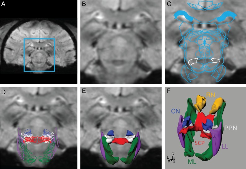
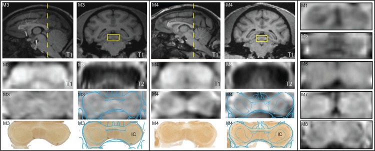
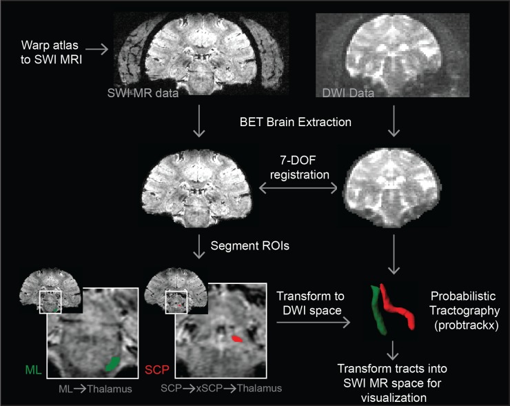
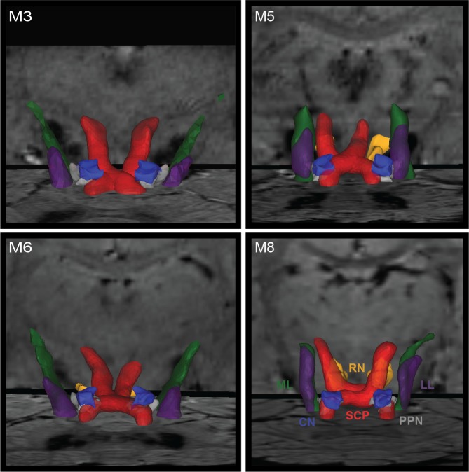
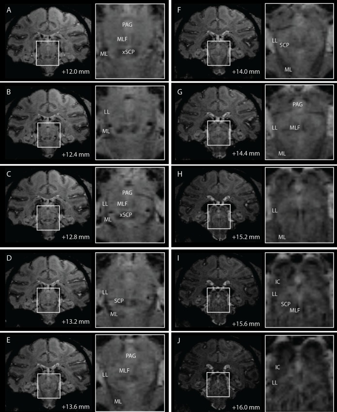
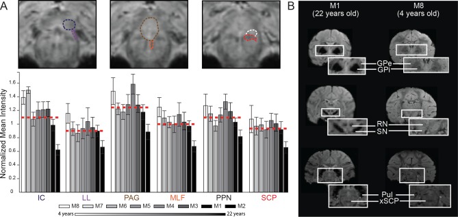
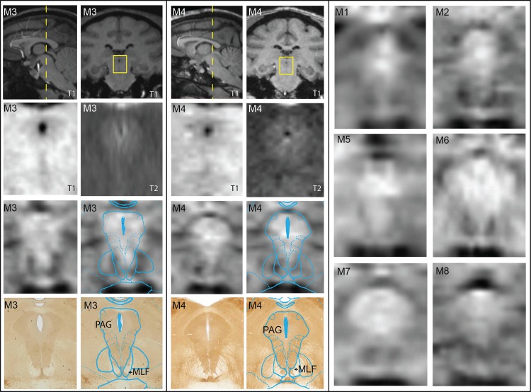
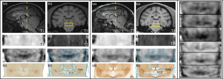
Structural brain imaging provides a critical framework for performing stereotactic and intraoperative MRI-guided surgical procedures, with procedural efficacy often dependent upon visualization of the target with which to operate. Here, we describe tools for in vivo, subject-specific visualization and demarcation of regions within the brainstem. High-field 7T susceptibility-weighted imaging and diffusion-weighted imaging of the brain were collected using a customized head coil from eight rhesus macaques. Fiber tracts including the superior cerebellar peduncle, medial lemniscus, and lateral lemniscus were identified using high-resolution probabilistic diffusion tractography, which resulted in three-dimensional fiber tract reconstructions that were comparable to those extracted from sequential application of a two-dimensional nonlinear brain atlas warping algorithm. In the susceptibility-weighted imaging, white matter tracts within the brainstem were also identified as hypointense regions, and the degree of hypointensity was age-dependent. This combination of imaging modalities also enabled identifying the location and extent of several brainstem nuclei, including the periaqueductal gray, pedunculopontine nucleus, and inferior colliculus. These clinically-relevant high-field imaging approaches have potential to enable more accurate and comprehensive subject-specific visualization of the brainstem and to ultimately improve patient-specific neurosurgical targeting procedures, including deep brain stimulation lead implantation.

摘要

脑结构成像为进行立体定向和术中磁共振成像引导的外科手术提供了关键框架,手术效果通常取决于对手术目标的可视化。在此,我们描述了用于在体、特定个体可视化和划分脑干内区域的工具。使用定制的头部线圈,对八只恒河猴进行了脑部的高场7T susceptibility加权成像和扩散加权成像。使用高分辨率概率扩散束描记法识别包括上小脑脚、内侧丘系和外侧丘系在内的纤维束,从而得到与从二维非线性脑图谱扭曲算法的顺序应用中提取的三维纤维束重建结果相当的结果。在susceptibility加权成像中,脑干内的白质束也被识别为低信号区域,且低信号程度与年龄相关。这种成像方式的组合还能够识别包括导水管周围灰质、脚桥核和下丘在内的几个脑干核团的位置和范围。这些与临床相关的高场成像方法有可能实现更准确、更全面的特定个体脑干可视化,并最终改善针对患者的神经外科靶向手术,包括深部脑刺激电极植入。

https://cdn.ncbi.nlm.nih.gov/pmc/blobs/eca4/4428864/dada00e56f71/pone.0127049.g008.jpg
https://cdn.ncbi.nlm.nih.gov/pmc/blobs/eca4/4428864/56efc117e30d/pone.0127049.g001.jpg
https://cdn.ncbi.nlm.nih.gov/pmc/blobs/eca4/4428864/dce797a8f057/pone.0127049.g002.jpg
https://cdn.ncbi.nlm.nih.gov/pmc/blobs/eca4/4428864/5ba9844db978/pone.0127049.g003.jpg
https://cdn.ncbi.nlm.nih.gov/pmc/blobs/eca4/4428864/262eda4dfc62/pone.0127049.g004.jpg
https://cdn.ncbi.nlm.nih.gov/pmc/blobs/eca4/4428864/e37847a3df9c/pone.0127049.g005.jpg
https://cdn.ncbi.nlm.nih.gov/pmc/blobs/eca4/4428864/d75be67cba39/pone.0127049.g006.jpg
https://cdn.ncbi.nlm.nih.gov/pmc/blobs/eca4/4428864/91f7e77b14be/pone.0127049.g007.jpg
https://cdn.ncbi.nlm.nih.gov/pmc/blobs/eca4/4428864/dada00e56f71/pone.0127049.g008.jpg

相似文献

1
In Vivo 7T MRI of the Non-Human Primate Brainstem.非人类灵长类动物脑干的体内7T磁共振成像
PLoS One. 2015 May 12;10(5):e0127049. doi: 10.1371/journal.pone.0127049. eCollection 2015.
2
Multimodal 7T Imaging of Thalamic Nuclei for Preclinical Deep Brain Stimulation Applications.用于临床前深部脑刺激应用的丘脑核团多模态7T成像
Front Neurosci. 2016 Jun 10;10:264. doi: 10.3389/fnins.2016.00264. eCollection 2016.
3
A practical approach to in vivo high-resolution diffusion tensor imaging of rhesus monkeys on a 3-T human scanner.一种在3-T人体扫描仪上对恒河猴进行体内高分辨率扩散张量成像的实用方法。
Magn Reson Imaging. 2009 Apr;27(3):335-46. doi: 10.1016/j.mri.2008.07.004. Epub 2008 Sep 2.
4
A systematic evaluation of intraoperative white matter tract shift in pediatric epilepsy surgery using high-field MRI and probabilistic high angular resolution diffusion imaging tractography.使用高场磁共振成像和概率性高角分辨率扩散成像纤维束示踪技术对小儿癫痫手术中白质束移位进行系统评估。
J Neurosurg Pediatr. 2017 May;19(5):592-605. doi: 10.3171/2016.11.PEDS16312. Epub 2017 Mar 17.
5
High-resolution MRI and diffusion-weighted imaging of the human habenula at 7 tesla.7特斯拉下人类缰核的高分辨率磁共振成像和扩散加权成像
J Magn Reson Imaging. 2014 Apr;39(4):1018-26. doi: 10.1002/jmri.24252. Epub 2013 Nov 20.
6
Postmortem diffusion MRI of the human brainstem and thalamus for deep brain stimulator electrode localization.用于脑深部刺激电极定位的人脑干和丘脑的尸检扩散磁共振成像
Hum Brain Mapp. 2015 Aug;36(8):3167-78. doi: 10.1002/hbm.22836. Epub 2015 Jun 3.
7
Magnetic resonance diffusion tensor imaging for the pedunculopontine nucleus: proof of concept and histological correlation.脚桥核的磁共振扩散张量成像:概念验证及组织学相关性
Brain Struct Funct. 2017 Aug;222(6):2547-2558. doi: 10.1007/s00429-016-1356-0. Epub 2017 Mar 10.
8
Population-averaged macaque brain atlas with high-resolution ex vivo DTI integrated into in vivo space.人群平均恒河猴大脑图谱,具有高分辨率离体扩散张量成像与体内空间整合。
Brain Struct Funct. 2017 Dec;222(9):4131-4147. doi: 10.1007/s00429-017-1463-6. Epub 2017 Jun 20.
9
Anatomical assessment of trigeminal nerve tractography using diffusion MRI: A comparison of acquisition b-values and single- and multi-fiber tracking strategies.使用扩散磁共振成像对三叉神经纤维束成像的解剖学评估:采集b值以及单纤维和多纤维追踪策略的比较
Neuroimage Clin. 2020;25:102160. doi: 10.1016/j.nicl.2019.102160. Epub 2020 Jan 8.
10
Track-Density Imaging of the Human Brainstem for Anatomic Localization of Fiber Tracts and Nerve Nuclei in Vivo: Initial Experience with 3-T Magnetic Resonance Imaging.用于体内纤维束和神经核团解剖定位的人脑干轨迹密度成像:3-T磁共振成像的初步经验
World Neurosurg. 2016 Sep;93:286-92. doi: 10.1016/j.wneu.2016.05.085. Epub 2016 Jun 4.

引用本文的文献

1
Direct localization and delineation of human pedunculopontine nucleus based on a self-supervised magnetic resonance image super-resolution method.基于自监督磁共振图像超分辨率方法的人类脑桥被盖脚核的直接定位和描绘。
Hum Brain Mapp. 2023 Jun 15;44(9):3781-3794. doi: 10.1002/hbm.26311. Epub 2023 Apr 25.
2
Evaluation of Rhesus Macaque Models for Cerebral Palsy.恒河猴脑性瘫痪模型的评估
Brain Sci. 2022 Sep 14;12(9):1243. doi: 10.3390/brainsci12091243.
3
Imaging and spectroscopic methods to investigate adult neurogenesis : New models and new avenues.

本文引用的文献

1
Mapping track density changes in nigrostriatal and extranigral pathways in Parkinson's disease.帕金森病中黑质纹状体和黑质外通路的追踪密度变化映射
Neuroimage. 2014 Oct 1;99:498-508. doi: 10.1016/j.neuroimage.2014.06.033. Epub 2014 Jun 20.
2
Cross-validation of serial optical coherence scanning and diffusion tensor imaging: a study on neural fiber maps in human medulla oblongata.连续光学相干扫描与扩散张量成像的交叉验证:一项关于人类延髓神经纤维图谱的研究。
Neuroimage. 2014 Oct 15;100:395-404. doi: 10.1016/j.neuroimage.2014.06.032. Epub 2014 Jun 20.
3
Spatial normalization of ultrahigh resolution 7 T magnetic resonance imaging data of the postmortem human subthalamic nucleus: a multistage approach.
用于研究成体神经发生的成像和光谱方法:新模型与新途径
Front Neurosci. 2022 Aug 5;16:933947. doi: 10.3389/fnins.2022.933947. eCollection 2022.
4
Ultra-high field (10.5T) diffusion-weighted MRI of the macaque brain.恒磁场强度为 10.5T 的猕猴大脑弥散加权磁共振成像。
Neuroimage. 2022 Jul 15;255:119200. doi: 10.1016/j.neuroimage.2022.119200. Epub 2022 Apr 13.
5
Post-Concussive Vestibular Dysfunction Is Related to Injury to the Inferior Vestibular Nerve.脑震荡后前庭功能障碍与下前庭神经损伤有关。
J Neurotrauma. 2022 Mar 7;39(11-12):829-40. doi: 10.1089/neu.2021.0447.
6
A 16-channel AC/DC array coil for anesthetized monkey whole-brain imaging at 7T.用于7T麻醉猴全脑成像的16通道交直流阵列线圈。
Neuroimage. 2020 Feb 15;207:116396. doi: 10.1016/j.neuroimage.2019.116396. Epub 2019 Nov 25.
7
Identification of the pedunculopontine nucleus and surrounding white matter tracts on 7T diffusion tensor imaging, combined with histological validation.在7T扩散张量成像上识别脚桥核及周围白质束,并结合组织学验证。
Surg Radiol Anat. 2019 Feb;41(2):187-196. doi: 10.1007/s00276-018-2120-3. Epub 2018 Oct 31.
8
Challenges and opportunities for brainstem neuroimaging with ultrahigh field MRI.超高场 MRI 用于脑干神经影像学的挑战与机遇。
Neuroimage. 2018 Mar;168:412-426. doi: 10.1016/j.neuroimage.2017.02.052. Epub 2017 Feb 21.
9
Depth-dependence of visual signals in the human superior colliculus at 9.4 T.9.4特斯拉下人类上丘视觉信号的深度依赖性
Hum Brain Mapp. 2017 Jan;38(1):574-587. doi: 10.1002/hbm.23404. Epub 2016 Sep 23.
10
Multimodal 7T Imaging of Thalamic Nuclei for Preclinical Deep Brain Stimulation Applications.用于临床前深部脑刺激应用的丘脑核团多模态7T成像
Front Neurosci. 2016 Jun 10;10:264. doi: 10.3389/fnins.2016.00264. eCollection 2016.
死后人类丘脑底核超高分辨率7T磁共振成像数据的空间归一化:一种多阶段方法。
Brain Struct Funct. 2015;220(3):1695-703. doi: 10.1007/s00429-014-0754-4. Epub 2014 Mar 25.
4
Quantifying inter-individual anatomical variability in the subcortex using 7 T structural MRI.利用 7T 结构 MRI 量化皮质下亚区的个体间解剖结构变异性。
Neuroimage. 2014 Jul 1;94:40-46. doi: 10.1016/j.neuroimage.2014.03.032. Epub 2014 Mar 17.
5
Fiber tractography of the axonal pathways linking the basal ganglia and cerebellum in Parkinson disease: implications for targeting in deep brain stimulation.帕金森病基底节和小脑之间轴突通路的纤维束追踪:对深部脑刺激靶点选择的影响。
J Neurosurg. 2014 Apr;120(4):988-96. doi: 10.3171/2013.12.JNS131537. Epub 2014 Jan 31.
6
High-resolution anatomy of the human brain stem using 7-T MRI: improved detection of inner structures and nerves?7T MRI 对人类脑干的高分辨率解剖:提高对内部结构和神经的检测?
Neuroradiology. 2014 Mar;56(3):177-86. doi: 10.1007/s00234-013-1312-0. Epub 2013 Dec 20.
7
Injury to a specific neural pathway detected by ultra-high-field MRI.超高场 MRI 检测到特定神经通路损伤。
Neurology. 2014 Jan 14;82(2):182-3. doi: 10.1212/WNL.0000000000000016. Epub 2013 Nov 27.
8
High-Resolution MR Imaging of the Human Brainstem In vivo at 7 Tesla.7 特斯拉人体脑干高分辨率磁共振成像。
Front Hum Neurosci. 2013 Oct 29;7:710. doi: 10.3389/fnhum.2013.00710. eCollection 2013.
9
New developments and applications of the MP2RAGE sequence--focusing the contrast and high spatial resolution R1 mapping.MP2RAGE 序列的新发展和应用——聚焦对比和高空间分辨率 R1 映射。
PLoS One. 2013 Jul 16;8(7):e69294. doi: 10.1371/journal.pone.0069294. Print 2013.
10
Computational modeling of pedunculopontine nucleus deep brain stimulation.脑桥被盖核深部脑刺激的计算建模。
J Neural Eng. 2013 Aug;10(4):045005. doi: 10.1088/1741-2560/10/4/045005. Epub 2013 May 31.