• 文献检索
  • 文档翻译
  • 深度研究
  • 学术资讯
  • Suppr Zotero 插件Zotero 插件
  • 邀请有礼
  • 套餐&价格
  • 历史记录
应用&插件
Suppr Zotero 插件Zotero 插件浏览器插件Mac 客户端Windows 客户端微信小程序
定价
高级版会员购买积分包购买API积分包
服务
文献检索文档翻译深度研究API 文档MCP 服务
关于我们
关于 Suppr公司介绍联系我们用户协议隐私条款
关注我们

Suppr 超能文献

核心技术专利:CN118964589B侵权必究
粤ICP备2023148730 号-1Suppr @ 2026

文献检索

告别复杂PubMed语法,用中文像聊天一样搜索,搜遍4000万医学文献。AI智能推荐,让科研检索更轻松。

立即免费搜索

文件翻译

保留排版,准确专业,支持PDF/Word/PPT等文件格式,支持 12+语言互译。

免费翻译文档

深度研究

AI帮你快速写综述,25分钟生成高质量综述,智能提取关键信息,辅助科研写作。

立即免费体验

利用磁共振成像(MRI)和乳腺X线摄影的综合信息识别乳腺癌

Identification of Breast Cancer Using Integrated Information from MRI and Mammography.

作者信息

Yang Shih-Neng, Li Fang-Jing, Liao Yen-Hsiu, Chen Yueh-Sheng, Shen Wu-Chung, Huang Tzung-Chi

机构信息

Department of Biomedical Imaging and Radiological Science, China Medical University, Taichung, Taiwan; Department of Radiation Oncology, China Medical University Hospital, Taichung, Taiwan.

Department of Biomedical Imaging and Radiological Science, China Medical University, Taichung, Taiwan.

出版信息

PLoS One. 2015 Jun 9;10(6):e0128404. doi: 10.1371/journal.pone.0128404. eCollection 2015.

DOI:10.1371/journal.pone.0128404
PMID:26056841
原文链接:https://pmc.ncbi.nlm.nih.gov/articles/PMC4461246/
Abstract

OBJECTIVES

Integration of information from corresponding regions between the breast MRI and an X-ray mammogram could benefit the detection of breast cancer in clinical diagnosis. We aimed to provide a framework of registration from breast MRI to mammography and to evaluate the diagnosis using the combined information.

MATERIALS AND METHODS

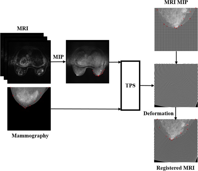
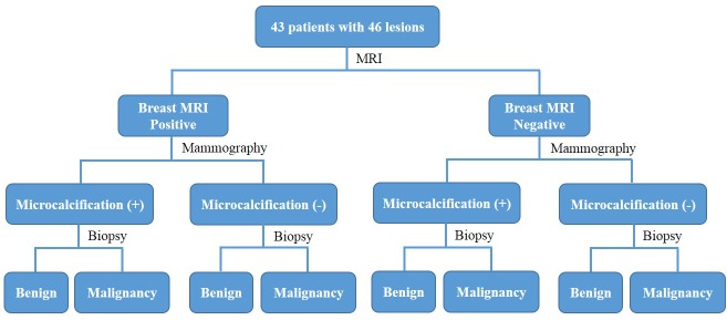
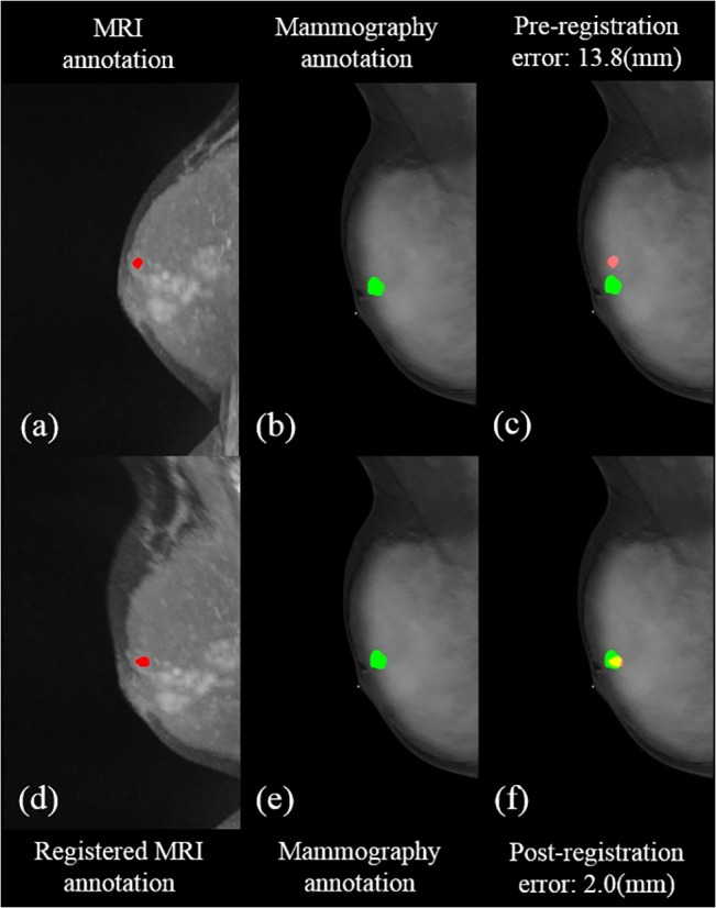
43 patients with 46 lesions underwent both MRI and mammography scans, and the interval between the two examinations was around one month. The distribution of malignant to benign lesions was 31/46 based on histological results. Maximum intensity projection and thin-plate spline methods were applied for image registration for MRI to mammography. The diagnosis using integrated information was evaluated using results of histology as the reference. The assessment of annotations and statistical analysis were performed by the two radiologists.

RESULTS

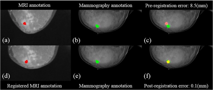
For the cranio-caudal view, the mean post-registration error between MRI and mammography was 2.2±1.9 mm. For the medio-lateral oblique view, the proposed approach performed even better with a mean error of 3.0±2.4 mm. In the diagnosis using MRI assessment with information of mammography, the sensitivity was 91.9±2.3% (29/31, 28/31), specificity 70.0±4.7% (11/15, 10/15), accuracy 84.8±3.1% (40/46, 38/46), positive predictive value 86.4±2.1% (29/33, 28/33) and negative predictive value 80.8±5.4% (11/13, 10/13).

CONCLUSION

MRI with the aid of mammography shows potential improvements of sensitivity, specificity, accuracy, PPV and NPV in clinical breast cancer diagnosis compared to the use of MRI alone.

摘要

目的

整合乳腺磁共振成像(MRI)与乳腺X线摄影相应区域的信息,有助于临床诊断中乳腺癌的检测。我们旨在提供一个从乳腺MRI到乳腺X线摄影的配准框架,并利用整合后的信息评估诊断效果。

材料与方法

43例患者共46个病灶接受了MRI和乳腺X线摄影扫描,两次检查间隔约1个月。根据组织学结果,恶性与良性病灶的分布为31/46。采用最大强度投影和薄板样条法对MRI与乳腺X线摄影进行图像配准。以组织学结果为参考,评估整合信息后的诊断效果。两位放射科医生进行标注评估和统计分析。

结果

在头尾位视图中,MRI与乳腺X线摄影配准后的平均误差为2.2±1.9毫米。在内外斜位视图中,所提出的方法表现更好,平均误差为3.0±2.4毫米。在利用乳腺X线摄影信息进行MRI评估的诊断中,敏感性为91.9±2.3%(29/31,28/31),特异性为70.0±4.7%(11/15,10/15),准确性为84.8±3.1%(40/46,38/46),阳性预测值为86.4±2.1%(29/33,28/33),阴性预测值为80.8±5.4%(11/13,10/13)。

结论

与单独使用MRI相比,借助乳腺X线摄影的MRI在临床乳腺癌诊断中显示出敏感性、特异性、准确性、阳性预测值和阴性预测值的潜在提高。

https://cdn.ncbi.nlm.nih.gov/pmc/blobs/17cc/4461246/3294ced8b162/pone.0128404.g005.jpg
https://cdn.ncbi.nlm.nih.gov/pmc/blobs/17cc/4461246/71d352051df4/pone.0128404.g001.jpg
https://cdn.ncbi.nlm.nih.gov/pmc/blobs/17cc/4461246/74cebb610e81/pone.0128404.g002.jpg
https://cdn.ncbi.nlm.nih.gov/pmc/blobs/17cc/4461246/68c6c373f942/pone.0128404.g003.jpg
https://cdn.ncbi.nlm.nih.gov/pmc/blobs/17cc/4461246/9f4ceee28e7c/pone.0128404.g004.jpg
https://cdn.ncbi.nlm.nih.gov/pmc/blobs/17cc/4461246/3294ced8b162/pone.0128404.g005.jpg

相似文献

1
Identification of Breast Cancer Using Integrated Information from MRI and Mammography.利用磁共振成像(MRI)和乳腺X线摄影的综合信息识别乳腺癌
PLoS One. 2015 Jun 9;10(6):e0128404. doi: 10.1371/journal.pone.0128404. eCollection 2015.
2
Contrast-enhanced MR imaging in patients with BI-RADS 3-5 microcalcifications.BI-RADS 3-5级微钙化患者的对比增强磁共振成像
Radiol Med. 2007 Mar;112(2):272-86. doi: 10.1007/s11547-007-0141-9. Epub 2007 Mar 19.
3
The challenge of imaging dense breast parenchyma: is magnetic resonance mammography the technique of choice? A comparative study with x-ray mammography and whole-breast ultrasound.致密乳腺实质成像的挑战:磁共振乳腺造影是首选技术吗?一项与乳腺X线摄影和全乳超声的对比研究。
Invest Radiol. 2009 Jul;44(7):412-21. doi: 10.1097/RLI.0b013e3181a53654.
4
Positron Emission Tomography/Magnetic Resonance Imaging for Local Tumor Staging in Patients With Primary Breast Cancer: A Comparison With Positron Emission Tomography/Computed Tomography and Magnetic Resonance Imaging.正电子发射断层扫描/磁共振成像用于原发性乳腺癌患者的局部肿瘤分期:与正电子发射断层扫描/计算机断层扫描及磁共振成像的比较
Invest Radiol. 2015 Aug;50(8):505-13. doi: 10.1097/RLI.0000000000000197.
5
Diagnostic accuracy of high-resolution MRI using a microscopy coil for patients with presumed DCIS following mammography screening.使用显微镜线圈的高分辨率MRI对乳腺钼靶筛查后疑似导管原位癌患者的诊断准确性。
J Magn Reson Imaging. 2007 Jan;25(1):96-103. doi: 10.1002/jmri.20809.
6
Breast lesion detection and characterization at contrast-enhanced MR mammography: gadobenate dimeglumine versus gadopentetate dimeglumine.对比增强磁共振乳腺造影术对乳腺病变的检测与特征分析:钆贝葡胺与钆喷酸葡胺的比较
Radiology. 2005 Oct;237(1):45-56. doi: 10.1148/radiol.2371041369. Epub 2005 Aug 26.
7
Performance evaluation of breast cancer diagnosis with mammography, ultrasonography and magnetic resonance imaging.乳腺摄影、超声和磁共振成像在乳腺癌诊断中的性能评估。
J Xray Sci Technol. 2018;26(5):805-813. doi: 10.3233/XST-18388.
8
The Value of Quality-Assured Magnetic Resonance Imaging of the Breast for the Early Detection of Breast Cancer in Asymptomatic Women.质量保证的乳腺磁共振成像对无症状女性乳腺癌早期检测的价值
J Comput Assist Tomogr. 2018 Jan/Feb;42(1):1-5. doi: 10.1097/RCT.0000000000000650.
9
Magnetic resonance imaging of the breast prior to biopsy.活检前的乳腺磁共振成像。
JAMA. 2004 Dec 8;292(22):2735-42. doi: 10.1001/jama.292.22.2735.
10
Accuracy of clinical examination, digital mammogram, ultrasound, and MRI in determining postneoadjuvant pathologic tumor response in operable breast cancer patients.临床检查、数字乳腺 X 线摄影、超声和 MRI 对可手术乳腺癌患者新辅助化疗后病理肿瘤反应的判断准确性。
Ann Surg Oncol. 2011 Oct;18(11):3160-3. doi: 10.1245/s10434-011-1919-5. Epub 2011 Sep 27.

引用本文的文献

1
Improving signal-to-noise ratio by maximal convolution of longitudinal and transverse magnetization components in MRI: application to the breast cancer detection.通过在 MRI 中对纵向和横向磁化分量进行最大卷积来提高信噪比:在乳腺癌检测中的应用。
Med Biol Eng Comput. 2024 Mar;62(3):941-954. doi: 10.1007/s11517-023-02994-w. Epub 2023 Dec 15.
2
Exploring the mechanism of resistance to vincristine in breast cancer cells using transcriptome sequencing technology.利用转录组测序技术探索乳腺癌细胞对长春新碱的耐药机制。
Oncol Lett. 2023 Oct 5;26(6):502. doi: 10.3892/ol.2023.14089. eCollection 2023 Dec.
3
Diagnosis of Breast Cancer Using Radiomics Models Built Based on Dynamic Contrast Enhanced MRI Combined With Mammography.

本文引用的文献

1
Evaluation of the role of dynamic contrast-enhanced MR imaging for patients with BI-RADS 3-4 microcalcifications.动态对比增强磁共振成像在BI-RADS 3-4级微钙化患者中的作用评估
PLoS One. 2014 Jun 13;9(6):e99669. doi: 10.1371/journal.pone.0099669. eCollection 2014.
2
Computer-aided evaluation as an adjunct to revised BI-RADS Atlas: improvement in positive predictive value at screening breast MRI.计算机辅助评估作为修订后的BI-RADS图谱的辅助手段:提高乳腺MRI筛查的阳性预测值
Eur Radiol. 2014 Aug;24(8):1800-7. doi: 10.1007/s00330-014-3166-1. Epub 2014 May 2.
3
MRI to X-ray mammography intensity-based registration with simultaneous optimisation of pose and biomechanical transformation parameters.
基于动态对比增强磁共振成像联合乳腺钼靶构建的影像组学模型用于乳腺癌诊断
Front Oncol. 2021 Nov 17;11:774248. doi: 10.3389/fonc.2021.774248. eCollection 2021.
4
Implementing Multilabeling, ADASYN, and ReliefF Techniques for Classification of Breast Cancer Diagnostic through Machine Learning: Efficient Computer-Aided Diagnostic System.通过机器学习实现多标记、ADASYN 和 ReliefF 技术对乳腺癌诊断的分类:高效的计算机辅助诊断系统。
J Healthc Eng. 2021 Mar 22;2021:5577636. doi: 10.1155/2021/5577636. eCollection 2021.
5
Converging blockchain and next-generation artificial intelligence technologies to decentralize and accelerate biomedical research and healthcare.融合区块链和下一代人工智能技术,以实现生物医学研究和医疗保健的去中心化并加速其发展。
Oncotarget. 2017 Nov 9;9(5):5665-5690. doi: 10.18632/oncotarget.22345. eCollection 2018 Jan 19.
6
Magnetic resonance mammography in comparison with mammography in the discovery of multifocal, multicentric and bilateral lesions of breast cancer.磁共振乳腺成像与乳腺X线摄影在发现乳腺癌多灶、多中心及双侧病变方面的比较。
Electron Physician. 2017 Aug 1;9(8):5083-5087. doi: 10.19082/5083. eCollection 2017 Aug.
7
Statistical analyses in trials for the comprehensive understanding of organogenesis and histogenesis in humans and mice.用于全面了解人类和小鼠器官发生及组织发生的试验中的统计分析。
J Biochem. 2016 Jun;159(6):553-61. doi: 10.1093/jb/mvw020. Epub 2016 Mar 2.
基于强度的 MRI 到 X 射线乳房摄影配准,同时优化姿态和生物力学变换参数。
Med Image Anal. 2014 May;18(4):674-83. doi: 10.1016/j.media.2014.03.003. Epub 2014 Mar 27.
4
Mammographic screening and reporting: a need for standardisation. A review.乳腺钼靶筛查与报告:标准化的必要性。综述。
Niger Postgrad Med J. 2013 Dec;20(4):346-51.
5
Diagnosis of breast masses from dynamic contrast-enhanced and diffusion-weighted MR: a machine learning approach.基于动态对比增强和弥散加权磁共振的乳腺肿块诊断:一种机器学习方法。
PLoS One. 2014 Jan 31;9(1):e87387. doi: 10.1371/journal.pone.0087387. eCollection 2014.
6
Automatic multimodal 2D/3D breast image registration using biomechanical FEM models and intensity-based optimization.基于生物力学有限元模型和基于强度的优化的自动多模态 2D/3D 乳房图像配准。
Med Image Anal. 2013 Feb;17(2):209-18. doi: 10.1016/j.media.2012.10.003. Epub 2012 Nov 29.
7
MRI to X-ray mammography registration using a volume-preserving affine transformation.MRI 到 X 射线乳房 X 光摄影配准使用体积保持仿射变换。
Med Image Anal. 2012 Jul;16(5):966-75. doi: 10.1016/j.media.2012.03.001. Epub 2012 Mar 28.
8
Nonmasslike enhancement at breast MR imaging: the added value of mammography and US for lesion categorization.乳腺磁共振成像中非肿块样强化:乳腺摄影和超声在病变分类中的附加价值。
Radiology. 2011 Oct;261(1):69-79. doi: 10.1148/radiol.11110190. Epub 2011 Jul 19.
9
2D/3D image fusion of X-ray mammograms with breast MRI: visualizing dynamic contrast enhancement in mammograms.X 射线乳房 X 光片与乳房 MRI 的 2D/3D 图像融合:在乳房 X 光片中可视化动态对比增强。
Int J Comput Assist Radiol Surg. 2012 May;7(3):339-48. doi: 10.1007/s11548-011-0623-z. Epub 2011 Jun 5.
10
Background parenchymal enhancement at breast MR imaging and breast cancer risk.背景实质增强在乳腺磁共振成像和乳腺癌风险中的作用。
Radiology. 2011 Jul;260(1):50-60. doi: 10.1148/radiol.11102156. Epub 2011 Apr 14.