Kinnear Ekaterina, Caproni Lisa J, Tregoning John S
Mucosal Infection & Immunity Group, Section of Virology, Imperial College London, St Mary's Campus, London, United Kingdom.
Touchlight Genetics Ltd., Leatherhead, Surrey, United Kingdom.
PLoS One. 2015 Jun 19;10(6):e0130375. doi: 10.1371/journal.pone.0130375. eCollection 2015.
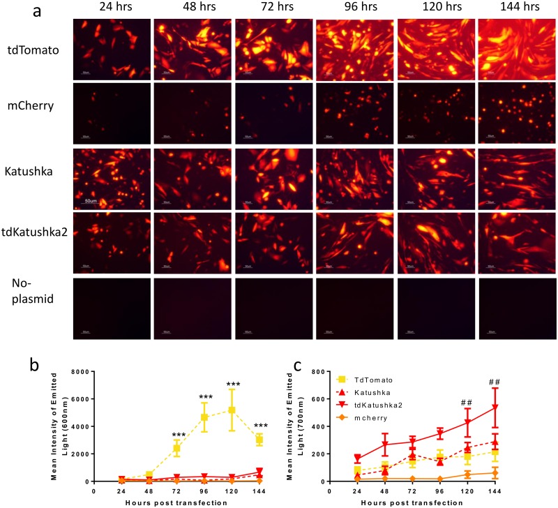
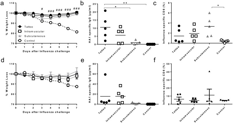
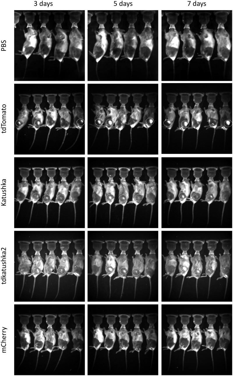
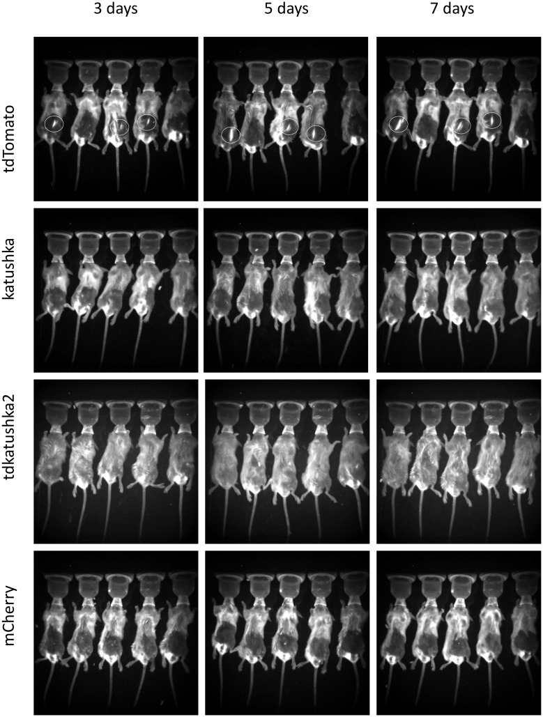
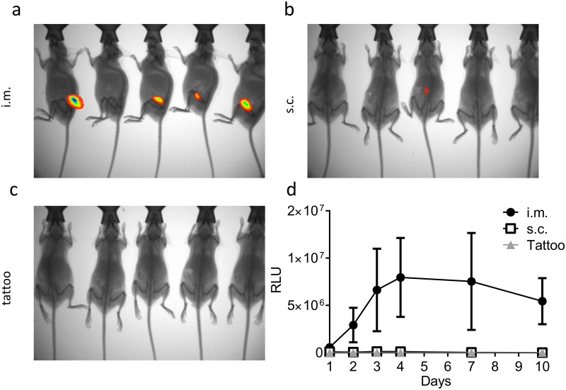
DNA vaccines can be manufactured cheaply, easily and rapidly and have performed well in pre-clinical animal studies. However, clinical trials have so far been disappointing, failing to evoke a strong immune response, possibly due to poor antigen expression. To improve antigen expression, improved technology to monitor DNA vaccine transfection efficiency is required. In the current study, we compared plasmid encoded tdTomato, mCherry, Katushka, tdKatushka2 and luciferase as reporter proteins for whole animal in vivo imaging. The intramuscular, subcutaneous and tattooing routes were compared and electroporation was used to enhance expression. We observed that overall, fluorescent proteins were not a good tool to assess expression from DNA plasmids, with a highly heterogeneous response between animals. Of the proteins used, intramuscular delivery of DNA encoding either tdTomato or luciferase gave the clearest signal, with some Katushka and tdKatushka2 signal observed. Subcutaneous delivery was weakly visible and nothing was observed following DNA tattooing. DNA encoding haemagglutinin was used to determine whether immune responses mirrored visible expression levels. A protective immune response against H1N1 influenza was induced by all routes, even after a single dose of DNA, though qualitative differences were observed, with tattooing leading to high antibody responses and subcutaneous DNA leading to high CD8 responses. We conclude that of the reporter proteins used, expression from DNA plasmids can best be assessed using tdTomato or luciferase. But, the disconnect between visible expression level and immunogenicity suggests that in vivo whole animal imaging of fluorescent proteins has limited utility for predicting DNA vaccine efficacy.
DNA疫苗可以低成本、轻松且快速地生产,并且在临床前动物研究中表现良好。然而,迄今为止的临床试验结果令人失望,未能引发强烈的免疫反应,这可能是由于抗原表达不佳所致。为了提高抗原表达,需要改进技术来监测DNA疫苗的转染效率。在本研究中,我们比较了质粒编码的tdTomato、mCherry、Katushka、tdKatushka2和荧光素酶作为报告蛋白,用于全动物体内成像。比较了肌肉内、皮下和纹身途径,并使用电穿孔来增强表达。我们观察到,总体而言,荧光蛋白不是评估DNA质粒表达的良好工具,不同动物之间的反应高度异质。在所使用的蛋白中,肌肉内递送编码tdTomato或荧光素酶的DNA产生了最清晰的信号,也观察到了一些Katushka和tdKatushka2信号。皮下递送信号较弱,DNA纹身后未观察到信号。编码血凝素的DNA用于确定免疫反应是否反映可见表达水平。所有途径均诱导了针对H1N1流感的保护性免疫反应,即使在单剂量DNA之后也是如此,尽管观察到了定性差异,纹身导致高抗体反应,皮下DNA导致高CD8反应。我们得出结论,在所使用的报告蛋白中,使用tdTomato或荧光素酶可以最好地评估DNA质粒的表达。但是,可见表达水平与免疫原性之间的脱节表明,荧光蛋白的体内全动物成像在预测DNA疫苗疗效方面的效用有限。