Bhat Vikas, Olmer Merissa, Joshi Shweta, Durden Donald L, Cramer Thomas J, Barnes Richard Fw, Ball Scott T, Hughes Tudor H, Silva Mauricio, Luck James V, Moore Randy E, Mosnier Laurent O, von Drygalski Annette
Department of Molecular and Experimental Medicine, The Scripps Research Institute, La Jolla, California.
Department of Medicine, University of California San Diego, San Diego, California.
Am J Hematol. 2015 Nov;90(11):1027-35. doi: 10.1002/ajh.24133. Epub 2015 Oct 8.
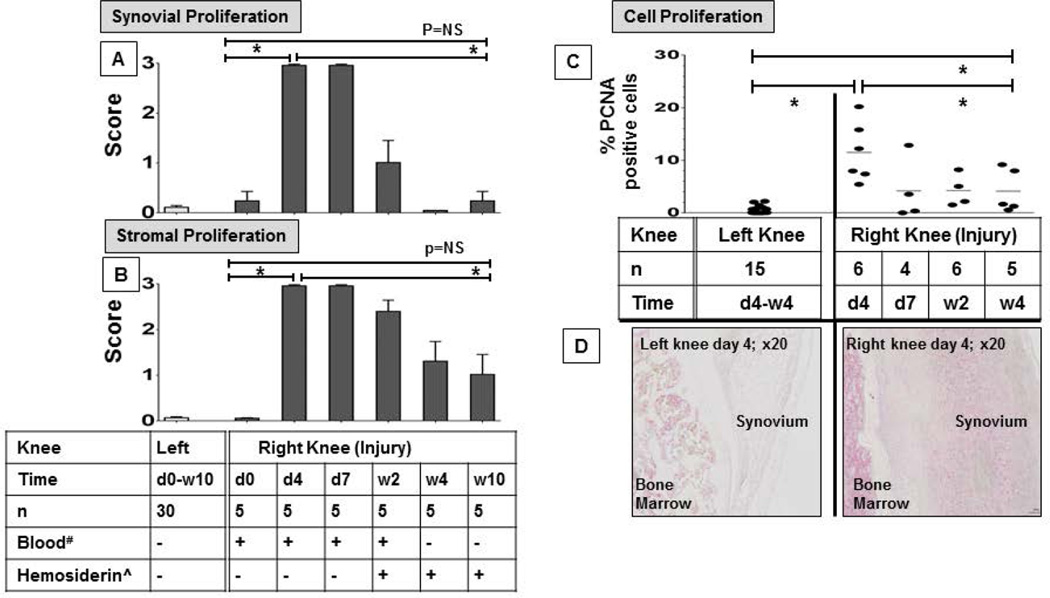
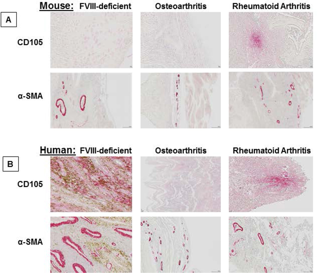
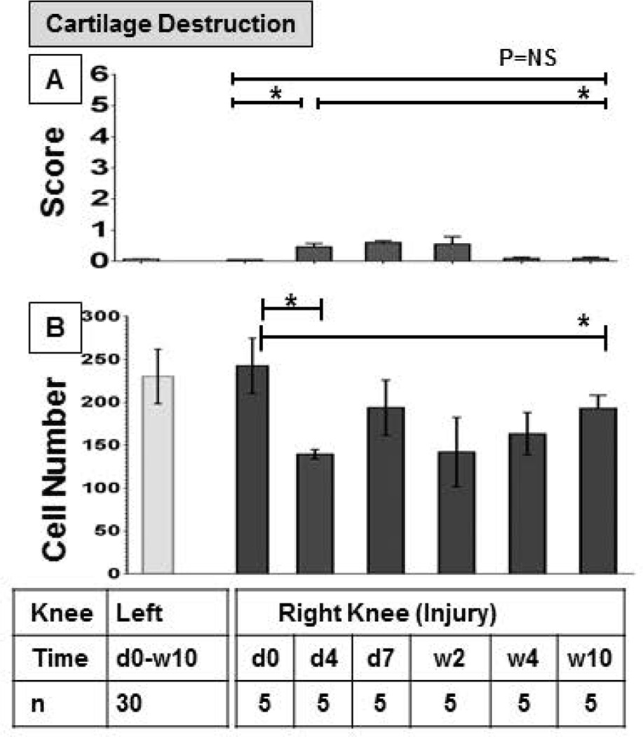
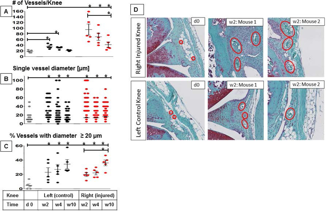
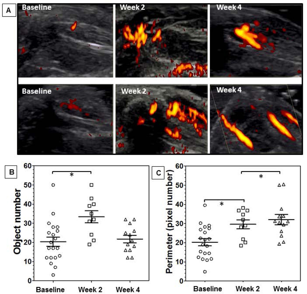
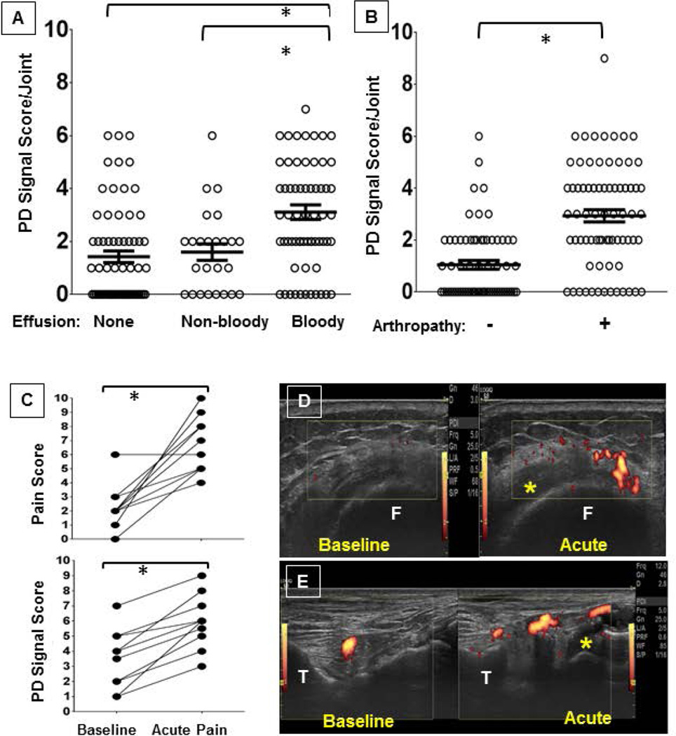
Hemophilic arthropathy is a debilitating condition that can develop as a consequence of frequent joint bleeding despite adequate clotting factor replacement. The mechanisms leading to repeated spontaneous bleeding are unknown. We investigated synovial, vascular, stromal, and cartilage changes in response to a single induced hemarthrosis in the FVIII-deficient mouse. We found soft-tissue hyperproliferation with marked induction of neoangiogenesis and evolving abnormal vascular architecture. While soft-tissue changes were rapidly reversible, abnormal vascularity persisted for months and, surprisingly, was also seen in uninjured joints. Vascular changes in FVIII-deficient mice involved pronounced remodeling with expression of α-Smooth Muscle Actin (SMA), Endoglin (CD105), and vascular endothelial growth factor, as well as alterations of joint perfusion as determined by in vivo imaging. Vascular architecture changes and pronounced expression of α-SMA appeared unique to hemophilia, as these were not found in joint tissue obtained from mouse models of rheumatoid arthritis and osteoarthritis and from patients with the same conditions. Evidence that vascular changes in hemophilia were significantly associated with bleeding and joint deterioration was obtained prospectively by dynamic in vivo imaging with musculoskeletal ultrasound and power Doppler of 156 joints (elbows, knees, and ankles) in a cohort of 26 patients with hemophilia at baseline and during painful episodes. These observations support the hypothesis that vascular remodeling contributes significantly to bleed propagation and development of hemophilic arthropathy. Based on these findings, the development of molecular targets for angiogenesis inhibition may be considered in this disease.
血友病性关节病是一种使人衰弱的病症,尽管进行了充分的凝血因子替代治疗,但仍可能因频繁的关节出血而发生。导致反复自发性出血的机制尚不清楚。我们研究了F VIII缺陷小鼠单次诱导关节积血后滑膜、血管、基质和软骨的变化。我们发现软组织过度增殖,伴有明显的新生血管形成诱导和不断演变的异常血管结构。虽然软组织变化可迅速逆转,但异常血管状态持续数月,令人惊讶的是,在未受伤的关节中也可见到。F VIII缺陷小鼠的血管变化涉及明显的重塑,伴有α-平滑肌肌动蛋白(SMA)、内皮糖蛋白(CD105)和血管内皮生长因子的表达,以及通过体内成像确定的关节灌注改变。血管结构变化和α-SMA的明显表达似乎是血友病所特有的,因为在类风湿性关节炎和骨关节炎小鼠模型以及患有相同病症的患者的关节组织中未发现这些变化。通过对26例血友病患者队列中156个关节(肘部、膝盖和脚踝)在基线和疼痛发作期间进行肌肉骨骼超声和功率多普勒动态体内成像,前瞻性地获得了血友病血管变化与出血和关节恶化显著相关的证据。这些观察结果支持了血管重塑对血友病性关节病的出血传播和发展有显著贡献的假说。基于这些发现,在这种疾病中可以考虑开发抑制血管生成的分子靶点。