Yu Peng, Zhang Jing, Yu Shuchun, Luo Zhenzhong, Hua Fuzhou, Yuan Linhui, Zhou Zhidong, Liu Qin, Du Xiaohong, Chen Sisi, Zhang Lieliang, Xu Guohai
Department of Cardiology, the Second Affiliated Hospital of Nanchang University, Nanchang 330000, China.
Department of Anesthesiology, the Second Affiliated Hospital of Nanchang University, Nanchang 330000, China.
PLoS One. 2015 Aug 11;10(8):e0134666. doi: 10.1371/journal.pone.0134666. eCollection 2015.
Myocardial infarction leads to heart failure. Autophagy is excessively activated in myocardial ischemia/reperfusion (I/R) in rats. The aim of this study is to investigate whether the protection of sevoflurane postconditioning (SPC) in myocardial I/R is through restored impaired autophagic flux.
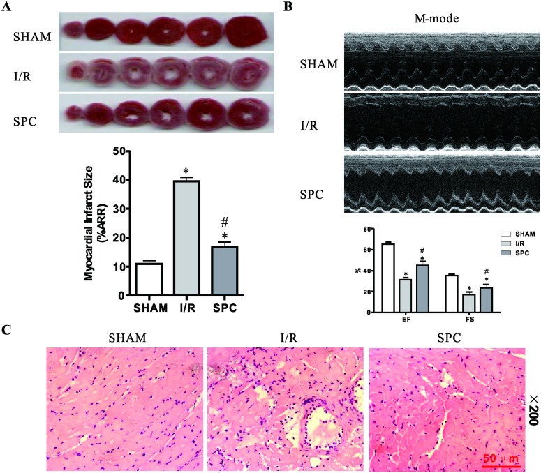
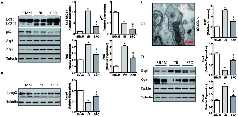
Except for the sham control (SHAM) group, each rat underwent 30 min occlusion of the left anterior descending coronary (LAD) followed by 2 h reperfusion. Cardiac infarction was determined by 2,3,5-triphenyltetrazolium chloride triazole (TTC) staining. Cardiac function was examined by hemodynamics and echocardiography. The activation of autophagy was evaluated by autophagosome accumulation, LC3 conversion and p62 degradation. Potential molecular mechanisms were investigated by immunoblotting, real-time PCR and immunofluorescence staining.
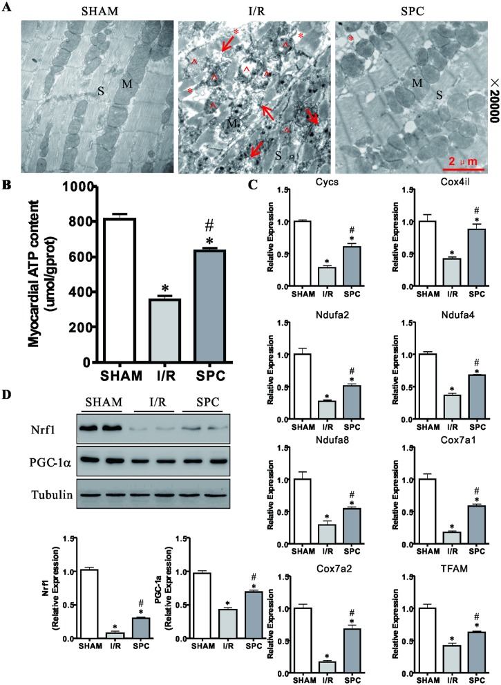
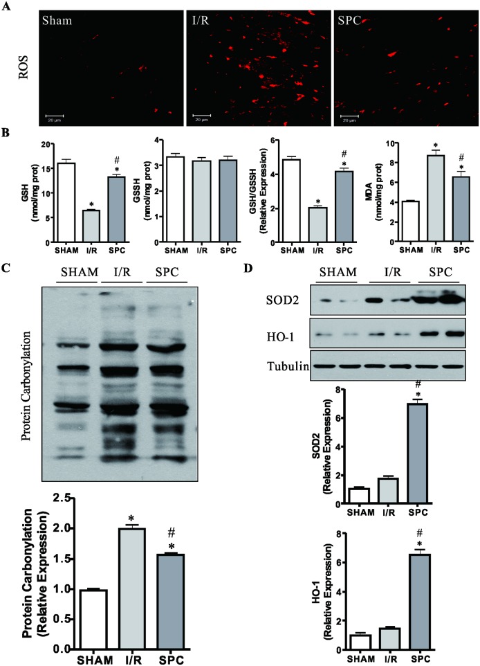
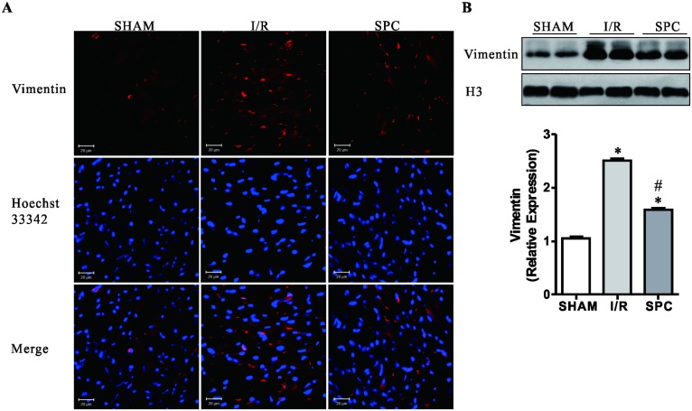
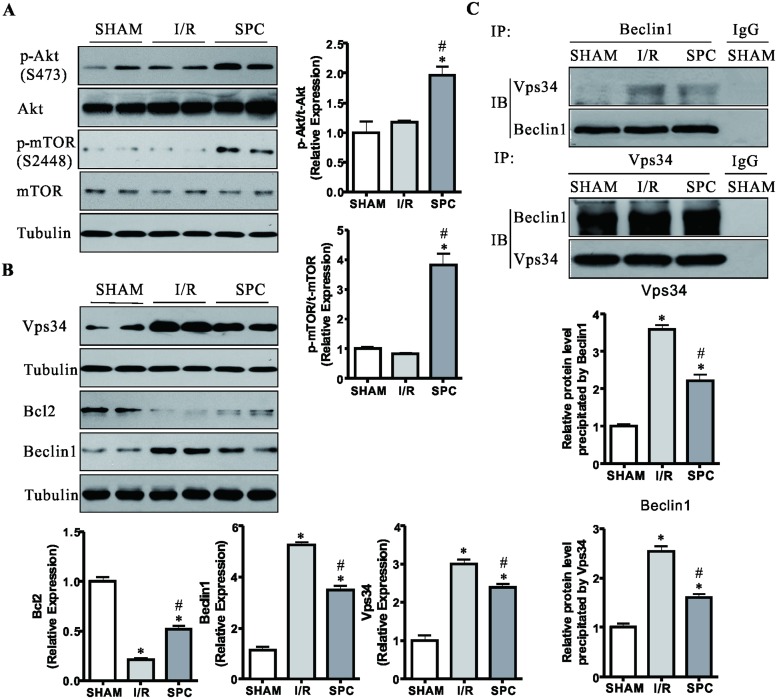
SPC improved the hemodynamic parameters, cardiac dysfunction, histopathological and ultrastructural damages, and decreased myocardial infarction size after myocardial I/R injury (P < 0.05 vs. I/R group). Compared with the cases in I/R group, myocardial ATP and NAD+ content, mitochondrial function related genes and proteins, and the expressions of SOD2 and HO-1 were increased, while the expressions of ROS and Vimentin were decreased in the SPC group (P < 0.05 vs. I/R group). SPC significantly activated Akt/mTOR signaling, and inhibited the formation of Vps34/Beclin1 complex via increasing expression of Bcl2 protein (P < 0.05 vs. I/R group). SPC suppressed elevated expressions of LC3 II/I ratio, Beclin1, Atg5 and Atg7 in I/R rat, which indicated that SPC inhibited over-activation of autophagy, and promoted autophagosome clearance. Meanwhile, SPC significantly suppressed the decline of Opa1 and increases of Drp1 and Parkin induced by I/R injury (P < 0.05 vs. I/R group). Moreover, SPC maintained the contents of ATP by reducing impaired mitochondria.
SPC protects rat hearts against I/R injury via ameliorating mitochondrial impairment, oxidative stress and rescuing autophagic clearance.
心肌梗死会导致心力衰竭。在大鼠心肌缺血/再灌注(I/R)过程中自噬被过度激活。本研究旨在探讨七氟醚后处理(SPC)对心肌I/R的保护作用是否是通过恢复受损的自噬通量来实现的。
除假手术对照组(SHAM)外,每只大鼠均接受左冠状动脉前降支(LAD)闭塞30分钟,随后再灌注2小时。通过2,3,5-氯化三苯基四氮唑(TTC)染色确定心肌梗死情况。通过血流动力学和超声心动图检查心功能。通过自噬体积累、LC3转化和p62降解评估自噬的激活情况。通过免疫印迹、实时PCR和免疫荧光染色研究潜在的分子机制。
SPC改善了心肌I/R损伤后的血流动力学参数、心脏功能障碍、组织病理学和超微结构损伤,并减小了心肌梗死面积(与I/R组相比,P<0.05)。与I/R组相比,SPC组心肌ATP和NAD+含量、线粒体功能相关基因和蛋白以及SOD2和HO-1的表达增加,而ROS和波形蛋白的表达降低(与I/R组相比,P<0.05)。SPC显著激活Akt/mTOR信号通路,并通过增加Bcl2蛋白表达抑制Vps34/Beclin1复合物的形成(与I/R组相比,P<0.05)。SPC抑制I/R大鼠中LC3 II/I比值、Beclin1、Atg5和Atg7表达的升高,这表明SPC抑制了自噬的过度激活,并促进了自噬体的清除。同时,SPC显著抑制I/R损伤诱导的Opa1下降以及Drp1和Parkin的增加(与I/R组相比,P<0.05)。此外,SPC通过减少受损线粒体来维持ATP含量。
SPC通过改善线粒体损伤、氧化应激和挽救自噬清除来保护大鼠心脏免受I/R损伤。