Hostettler Franziska C, Wiener Dominique J, Welle Monika M, Posthaus Horst, Geissbühler Urs
Clinical Radiology, Department of Clinical Veterinary Medicine, Vetsuisse Faculty, University of Bern, post office box 8466, CH-3001, Bern, Switzerland.
Institute of Animal Pathology, Vetsuisse Faculty, University of Bern, Länggassstrasse 122, CH-3012, Bern, Switzerland.
BMC Vet Res. 2015 Sep 2;11:229. doi: 10.1186/s12917-015-0544-0.
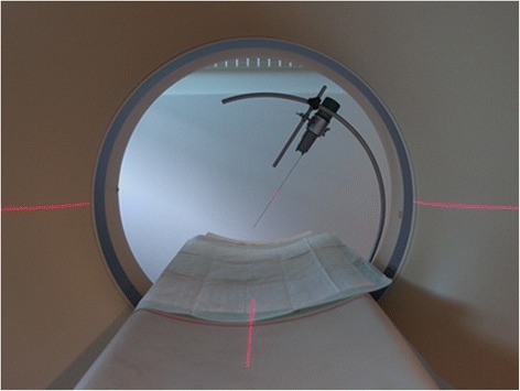
Bernese mountain dogs are reported to have a shorter life expectancy than other breeds. A major reason for this has been assigned to a high tumour prevalence, especially of histiocytic sarcoma. The efforts made by the breeding clubs to improve the longevity with the help of genetic tests and breeding value estimations are impeded by insufficiently reliable diagnoses regarding the cause of death. The current standard for post mortem examination in animals is performance of an autopsy. In human forensic medicine, imaging modalities, such as computed tomography and magnetic resonance imaging, are used with increasing frequency as a complement to autopsy. The present study investigates, whether post mortem computed tomography in combination with core needle biopsy is able to provide a definitive diagnosis of histiocytic sarcoma. For this purpose we have analysed the results of post mortem computed tomography and core needle biopsy in eleven Bernese mountain dogs. In the subsequent autopsy, every dog had a definitive diagnosis of histiocytic sarcoma, based on immunohistochemistry.
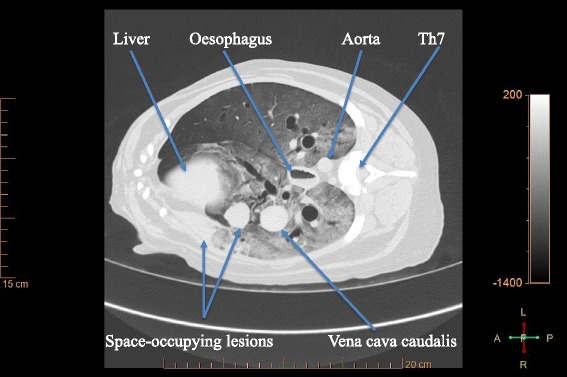
Computed tomography revealed space-occupying lesions in all dogs. Lesion detection by post mortem computed tomography was similar to lesion detection in autopsy for lung tissue (9 cases in computed tomography / 8 cases in autopsy), thoracic lymph nodes (9/8), spleen (6/7), kidney (2/2) and bone (3/3). Hepatic nodules, however, were difficult to detect with our scanning protocol (2/7). Histology of the core needle biopsies provided definitive diagnoses of histiocytic sarcoma in ten dogs, including confirmation by immunohistochemistry in six dogs. The biopsy samples of the remaining dog did not contain any identifiable neoplastic cells. Autolysis was the main reason for uncertain histological diagnoses.
Post mortem computed tomography is a fast and effective method for the detection of lesions suspicious for histiocytic sarcoma in pulmonary, thoracic lymphatic, splenic, osseous and renal tissue. Optimization of the procedure regarding the scanning protocol and tissue sample size and number will improve the accuracy of the method.
据报道,伯恩山犬的预期寿命比其他犬种短。造成这种情况的一个主要原因是肿瘤患病率高,尤其是组织细胞肉瘤。繁殖俱乐部借助基因检测和育种价值评估来提高犬只寿命的努力,因死因诊断可靠性不足而受到阻碍。目前动物尸检的标准做法是进行解剖。在人类法医学中,计算机断层扫描和磁共振成像等成像方式作为解剖的补充,使用频率越来越高。本研究调查死后计算机断层扫描结合粗针活检是否能够对组织细胞肉瘤做出明确诊断。为此,我们分析了11只伯恩山犬的死后计算机断层扫描和粗针活检结果。在随后的解剖中,每只犬均基于免疫组织化学确诊为组织细胞肉瘤。
计算机断层扫描显示所有犬均有占位性病变。死后计算机断层扫描对病变的检测与解剖时对肺组织(计算机断层扫描9例/解剖8例)、胸段淋巴结(9/8)、脾脏(6/7)、肾脏(2/2)和骨骼(3/3)的病变检测相似。然而,按照我们的扫描方案,肝结节很难检测到(2/7)。粗针活检的组织学检查对10只犬确诊为组织细胞肉瘤,其中6只犬经免疫组织化学证实。其余一只犬的活检样本未发现任何可识别的肿瘤细胞。自溶是组织学诊断不确定的主要原因。
死后计算机断层扫描是检测肺、胸段淋巴、脾、骨和肾组织中可疑组织细胞肉瘤病变的快速有效方法。优化扫描方案、组织样本大小和数量等操作将提高该方法的准确性。