Gutman David A, Dunn William D, Grossmann Patrick, Cooper Lee A D, Holder Chad A, Ligon Keith L, Alexander Brian M, Aerts Hugo J W L
Departments of Neurology, Emory University School of Medicine, Atlanta, GA, USA.
Biomedical Informatics, Emory University School of Medicine, 1648 Pierce Dr NE, Atlanta, GA, 30307, USA.
Neuroradiology. 2015 Dec;57(12):1227-37. doi: 10.1007/s00234-015-1576-7. Epub 2015 Sep 4.
MR imaging can noninvasively visualize tumor phenotype characteristics at the macroscopic level. Here, we investigated whether somatic mutations are associated with and can be predicted by MRI-derived tumor imaging features of glioblastoma (GBM).
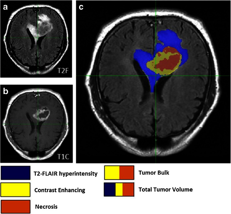
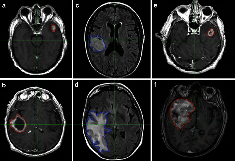
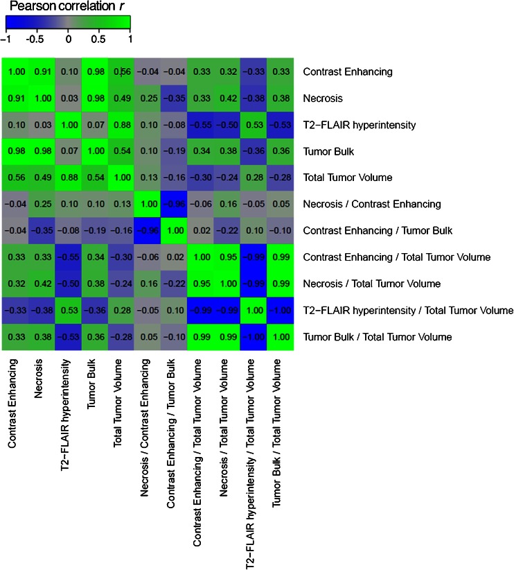
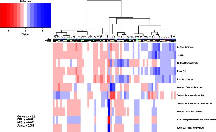
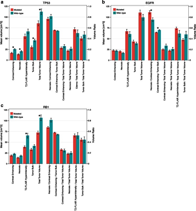
Seventy-six GBM patients were identified from The Cancer Imaging Archive for whom preoperative T1-contrast (T1C) and T2-FLAIR MR images were available. For each tumor, a set of volumetric imaging features and their ratios were measured, including necrosis, contrast enhancing, and edema volumes. Imaging genomics analysis assessed the association of these features with mutation status of nine genes frequently altered in adult GBM. Finally, area under the curve (AUC) analysis was conducted to evaluate the predictive performance of imaging features for mutational status.
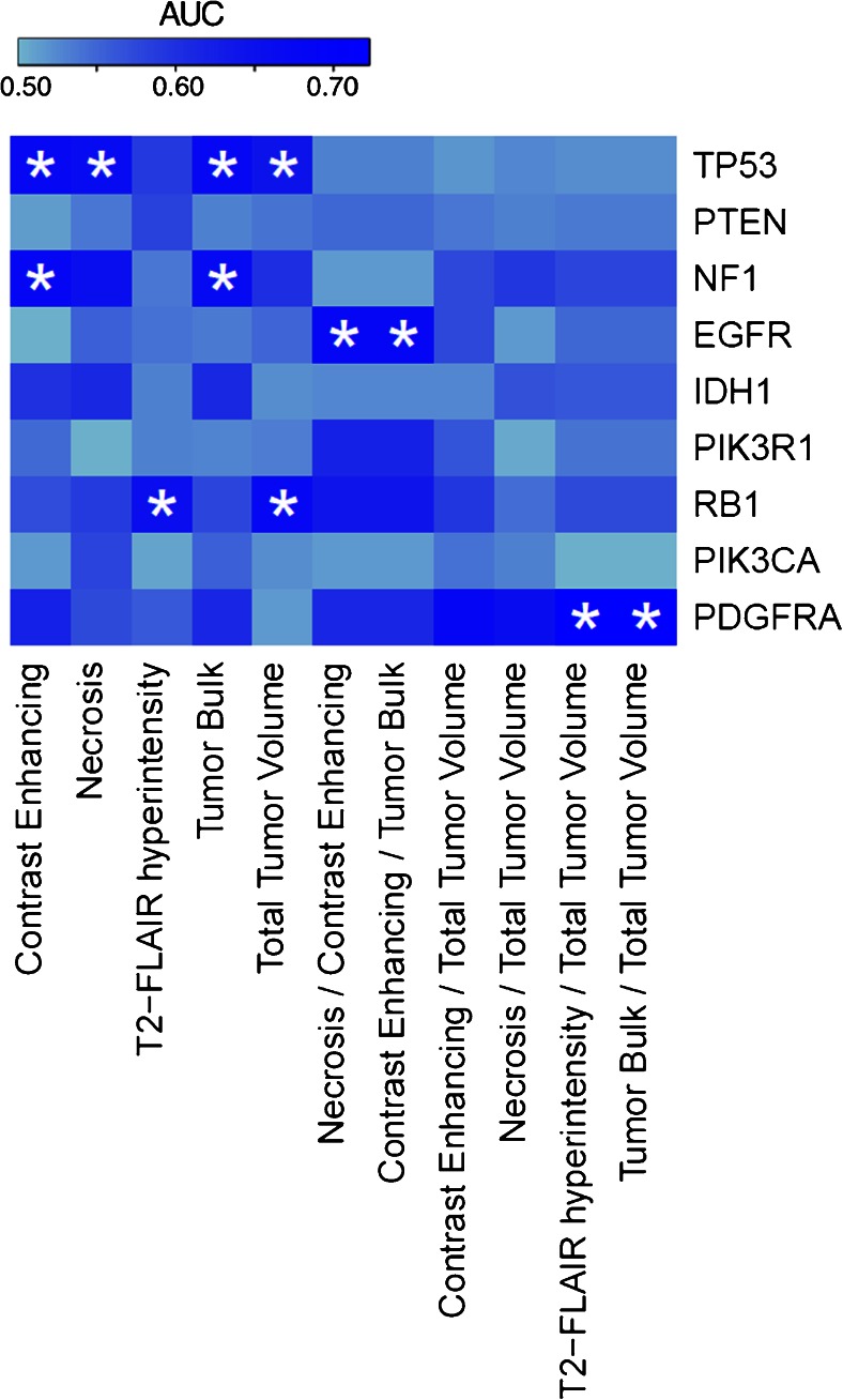
Our results demonstrate that MR imaging features are strongly associated with mutation status. For example, TP53-mutated tumors had significantly smaller contrast enhancing and necrosis volumes (p = 0.012 and 0.017, respectively) and RB1-mutated tumors had significantly smaller edema volumes (p = 0.015) compared to wild-type tumors. MRI volumetric features were also found to significantly predict mutational status. For example, AUC analysis results indicated that TP53, RB1, NF1, EGFR, and PDGFRA mutations could each be significantly predicted by at least one imaging feature.
MRI-derived volumetric features are significantly associated with and predictive of several cancer-relevant, drug-targetable DNA mutations in glioblastoma. These results may shed insight into unique growth characteristics of individual tumors at the macroscopic level resulting from molecular events as well as increase the use of noninvasive imaging in personalized medicine.
磁共振成像(MR)能够在宏观层面无创地显示肿瘤表型特征。在此,我们研究了体细胞突变是否与胶质母细胞瘤(GBM)的MRI衍生肿瘤成像特征相关,以及能否通过这些特征进行预测。
从癌症影像存档库中识别出76例GBM患者,这些患者术前有T1加权增强(T1C)和T2液体衰减反转恢复(T2-FLAIR)MR图像。对于每个肿瘤,测量一组体积成像特征及其比率,包括坏死、增强和水肿体积。影像基因组学分析评估了这些特征与成人GBM中九个经常发生改变的基因的突变状态之间的关联。最后,进行曲线下面积(AUC)分析,以评估成像特征对突变状态的预测性能。
我们的结果表明,MR成像特征与突变状态密切相关。例如,与野生型肿瘤相比,TP53突变的肿瘤增强和坏死体积显著更小(分别为p = 0.012和0.017),RB1突变的肿瘤水肿体积显著更小(p = 0.015)。还发现MRI体积特征能够显著预测突变状态。例如,AUC分析结果表明,TP53、RB1、NF1、EGFR和PDGFRA突变中的每一种都至少可由一种成像特征显著预测。
MRI衍生的体积特征与胶质母细胞瘤中几种与癌症相关的、可药物靶向的DNA突变显著相关并具有预测性。这些结果可能有助于深入了解分子事件导致的个体肿瘤在宏观层面的独特生长特征,以及增加无创成像在个性化医疗中的应用。