Zhou Jiawei, Baker Daniel H, Simard Mathieu, Saint-Amour Dave, Hess Robert F
McGill Vision Research, Department of Ophthalmology, McGill University, Montréal, Canada.
Department of Psychology, University of York, Heslington, York, UK.
Restor Neurol Neurosci. 2015;33(3):381-7. doi: 10.3233/RNN-140472.
Several recent studies have demonstrated that following short-term monocular deprivation in normal adults, the patched eye, rather than the unpatched eye, becomes stronger in subsequent binocular viewing. However, little is known about the site and nature of the underlying processes. In this study, we examine the underlying mechanisms by measuring steady-state visual evoked potentials (SSVEPs) as an index of the neural contrast response in early visual areas.
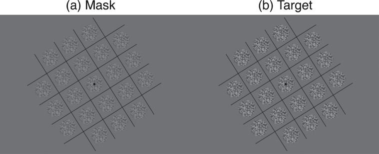
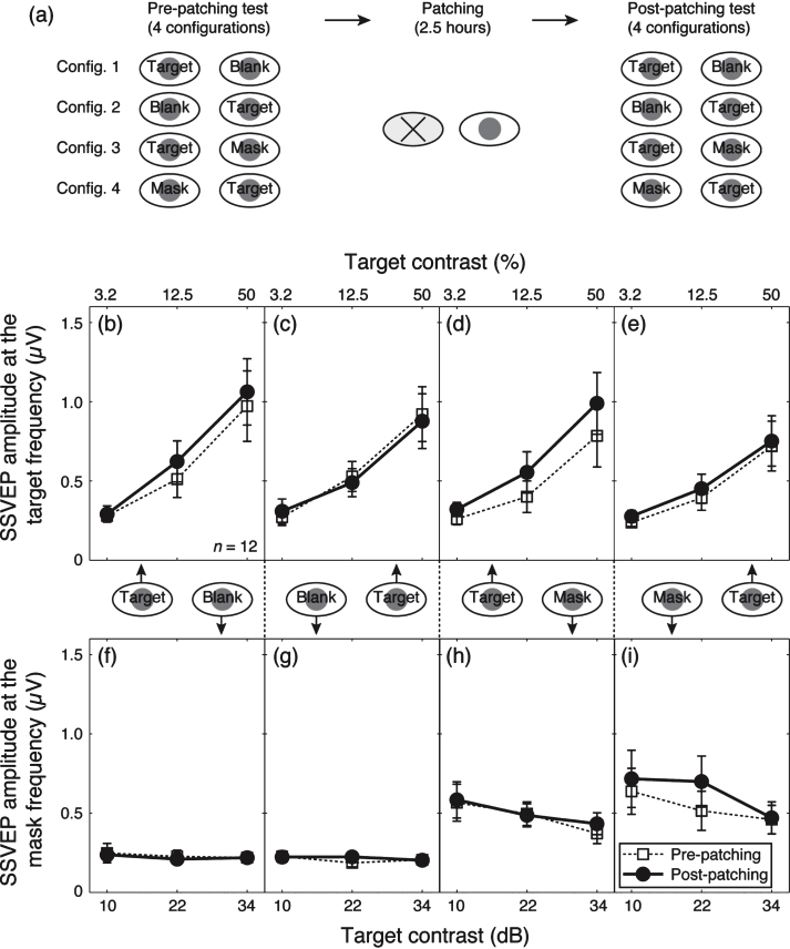
The experiment consisted of three consecutive stages: a pre-patching EEG recording (14 minutes), a monocular patching stage (2.5 hours) and a post-patching EEG recording (14 minutes; started immediately after the removal of the patch). During the patching stage, a diffuser (transmits light but not pattern) was placed in front of one randomly selected eye. During the EEG recording stage, contrast response functions for each eye were measured.
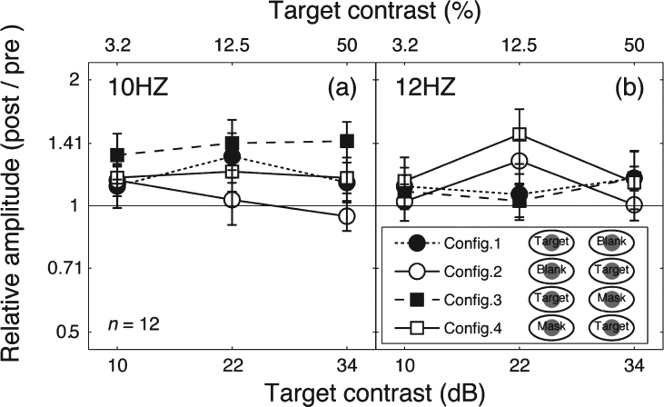
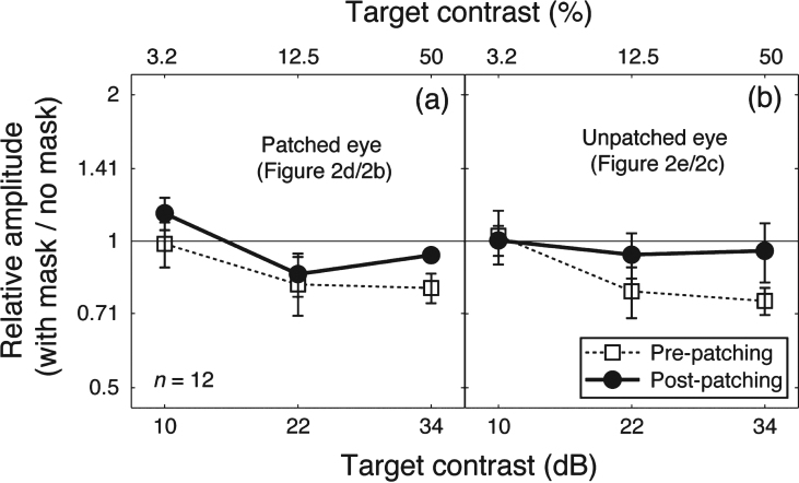
The neural responses from the patched eye increased after the removal of the patch, whilst the responses from the unpatched eye remained the same. Such phenomena occurred under both monocular and dichoptic viewing conditions.
We interpret this eye dominance plasticity in adult human visual cortex as homeostatic intrinsic plasticity regulated by an increase of contrast-gain in the patched eye.
最近的几项研究表明,正常成年人短期单眼剥夺后,在随后的双眼视觉中,被遮盖的眼睛而非未被遮盖的眼睛会变得更强。然而,对于其潜在过程的部位和性质知之甚少。在本研究中,我们通过测量稳态视觉诱发电位(SSVEP)作为早期视觉区域神经对比反应的指标,来研究其潜在机制。
实验包括三个连续阶段:遮盖前脑电图记录(14分钟)、单眼遮盖阶段(2.5小时)和遮盖后脑电图记录(14分钟;在去除眼罩后立即开始)。在遮盖阶段,将一个漫射器(透光但不透图案)放置在一只随机选择的眼睛前面。在脑电图记录阶段,测量每只眼睛的对比反应函数。
去除眼罩后,被遮盖眼睛的神经反应增加,而未被遮盖眼睛的反应保持不变。这种现象在单眼和双眼分视条件下均会出现。
我们将成年人类视觉皮层中的这种眼优势可塑性解释为由被遮盖眼睛的对比增益增加所调节的稳态内在可塑性。