Golestani N, EtehadTavakol M, Ng Eyk
Electrical and Computer Engineering Department, Isfahan University of Technology, Iran, Isfahan, 84154, Iran; e-mail:
Medical Image and Signal Processing Research Centre, Isfahan University of Medical Sciences, Isfahan 81745-319, Iran; e-mail:
EXCLI J. 2014 Mar 13;13:241-51. eCollection 2014.
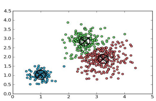
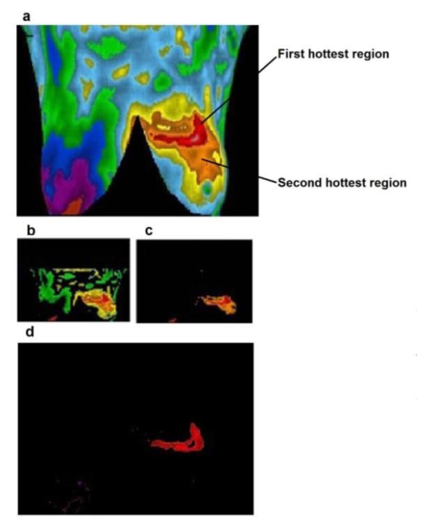
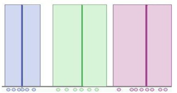
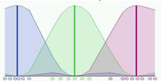
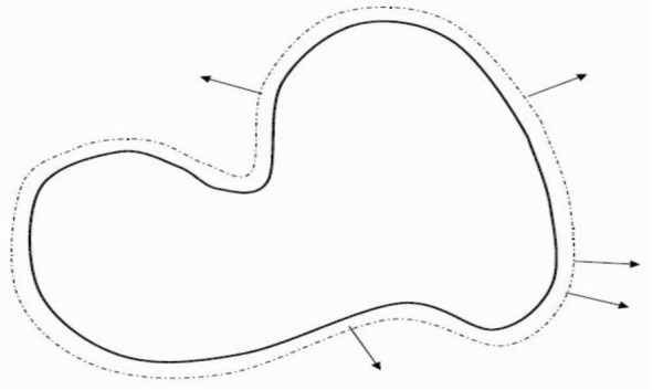
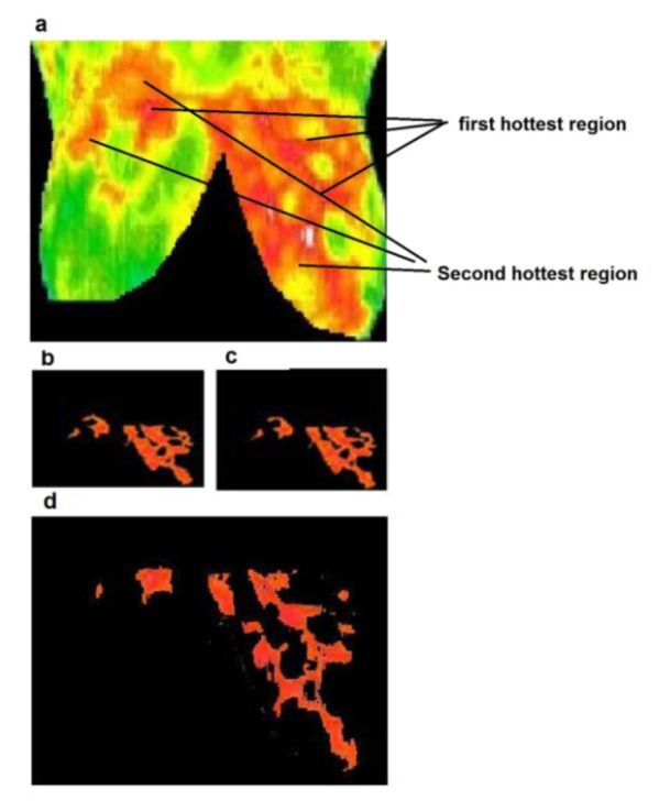
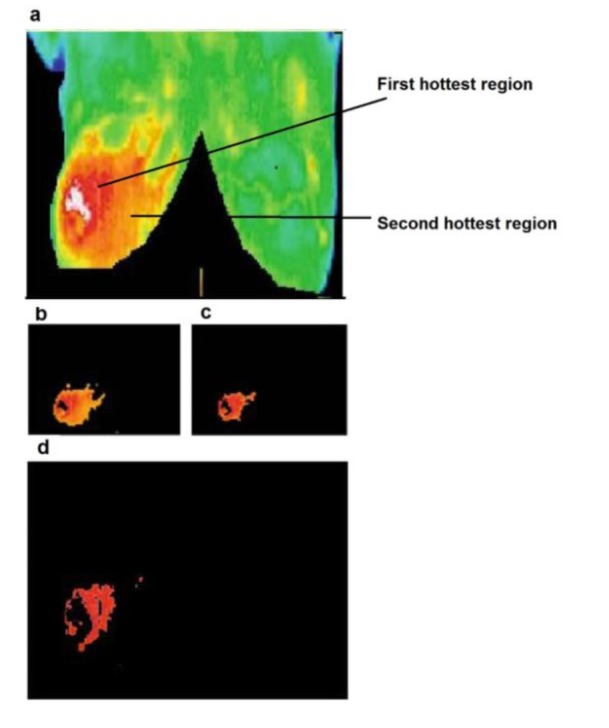
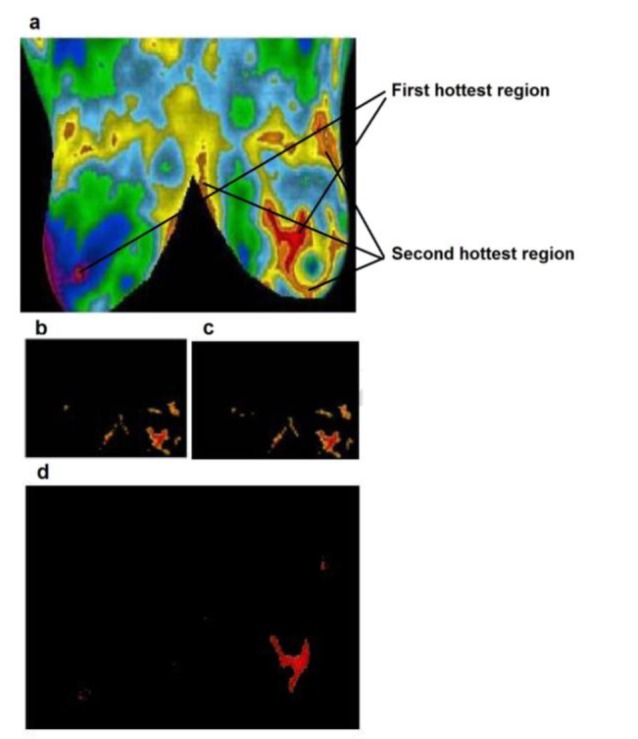
Breast thermography is a physiological test that provides information based on the temperature changes in breast. It records the temperature distribution of a body using the infrared radiation emitted by the surface of that body. Precancerous tissue and the area around a cancerous tumor have higher temperature due to angiogenesis, and higher chemical and blood vessel activity than a normal breast; hence breast thermography has potential to detect early abnormal changes in breast tissues. It can detect the first sign of forming up cancer before mammography can detect. The thermal information can be shown in a pseudo colored image where each color represents a specific range of temperature. Various methods can be applied to extract hot regions for detecting suspected regions of interests in the breast infrared images and potentially suspicious tissues. Image segmentation techniques can play an important role to segment and extract these regions in the breast infrared images. Shape, size and borders of the hottest regions of the images can help to determine features which are used to detect abnormalities. In this paper, three image segmentation methods: k-means, fuzzy c-means and level set are discussed and compared. These three methods are tested for different cases such as fibrocystic, inflammatory cancer cases. The hottest regions of thermal breast images in all cases are extracted and compared to the original images. According to the results, level set method is a more accurate approach and has potential to extract almost exact shape of tumors.
乳房热成像术是一种基于乳房温度变化提供信息的生理测试。它利用身体表面发出的红外辐射记录身体的温度分布。由于血管生成以及比正常乳房更高的化学和血管活性,癌前组织和癌性肿瘤周围区域的温度更高;因此,乳房热成像术有潜力检测乳房组织早期的异常变化。它能在乳房X线摄影术检测到之前,检测出癌症形成的首个迹象。热信息可以显示在伪彩色图像中,其中每种颜色代表特定的温度范围。可以应用各种方法来提取热点区域,以检测乳房红外图像中可疑的感兴趣区域以及潜在的可疑组织。图像分割技术在分割和提取乳房红外图像中的这些区域方面可以发挥重要作用。图像最热区域的形状、大小和边界有助于确定用于检测异常的特征。本文讨论并比较了三种图像分割方法:k均值法、模糊c均值法和水平集法。对这三种方法在不同病例(如纤维囊性、炎性癌病例)中进行了测试。提取了所有病例中乳房热图像的最热区域,并与原始图像进行比较。根据结果,水平集方法是一种更准确的方法,有潜力提取几乎精确的肿瘤形状。