Jann Kay, Hernandez Leanna M, Beck-Pancer Devora, McCarron Rosemary, Smith Robert X, Dapretto Mirella, Wang Danny J J
Laboratory of FMRI Technology (LOFT), Ahmanson-Lovelace Brain Mapping Center, Department of Neurology, University of California Los Angeles, California.
Department of Psychiatry and Biobehavioral Sciences, University of California Los Angeles, California.
Brain Behav. 2015 Sep;5(9):e00358. doi: 10.1002/brb3.358. Epub 2015 Jun 25.
Neuroimaging studies can shed light on the neurobiological underpinnings of autism spectrum disorders (ASD). Studies of the resting brain have shown both altered baseline metabolism from PET/SPECT and altered functional connectivity (FC) of intrinsic brain networks based on resting-state fMRI. To date, however, no study has investigated these two physiological parameters of resting brain function jointly, or explored the relationship between these measures and ASD symptom severity.
Here, we used pseudo-continuous arterial spin labeling with 3D background-suppressed GRASE to assess resting cerebral blood flow (CBF) and FC in 17 youth with ASD and 22 matched typically developing (TD) children.
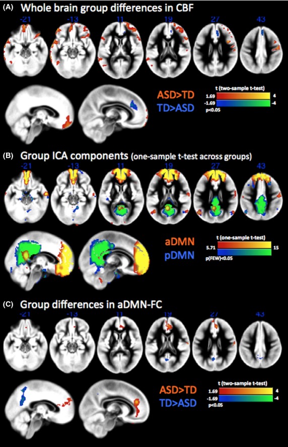
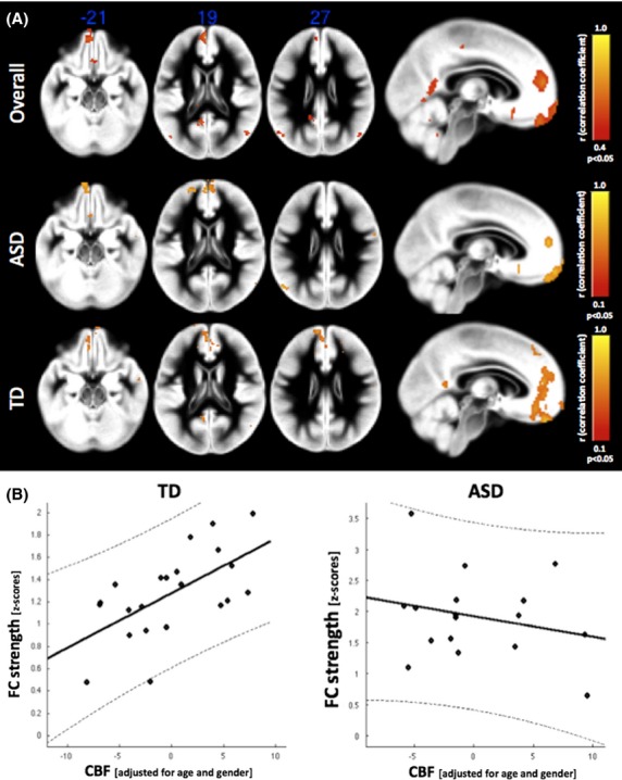
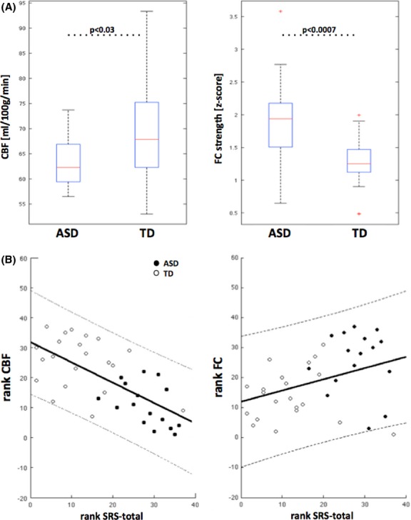
A pattern of altered resting perfusion was found in ASD versus TD children including frontotemporal hyperperfusion and hypoperfusion in the dorsal anterior cingulate cortex. We found increased local FC in the anterior module of the default mode network (DMN) accompanied by decreased CBF in the same area. In our cohort, both alterations were associated with greater social impairments as assessed with the Social Responsiveness Scale (SRS-total T scores). While FC was correlated with CBF in TD children, this association between FC and baseline perfusion was disrupted in children with ASD. Furthermore, there was reduced long-range FC between anterior and posterior modules of the DMN in children with ASD.
Taken together, the findings of this study--the first to jointly assess resting CBF and FC in ASD--highlight new avenues for identifying novel imaging markers of ASD symptomatology.
神经影像学研究有助于揭示自闭症谱系障碍(ASD)的神经生物学基础。静息态脑研究显示,正电子发射断层扫描/单光子发射计算机断层扫描(PET/SPECT)的基线代谢发生改变,基于静息态功能磁共振成像(fMRI)的脑内固有网络的功能连接(FC)也发生改变。然而,迄今为止,尚无研究同时调查静息态脑功能的这两个生理参数,或探究这些测量指标与ASD症状严重程度之间的关系。
在此,我们使用3D背景抑制梯度自旋回波伪连续动脉自旋标记技术,评估17名患有ASD的青少年和22名匹配的发育正常(TD)儿童的静息脑血流量(CBF)和FC。
与TD儿童相比,在ASD儿童中发现了静息灌注改变的模式,包括额颞叶灌注过多和背侧前扣带回皮质灌注不足。我们发现默认模式网络(DMN)前模块的局部FC增加,同时该区域的CBF减少。在我们的队列中,根据社会反应量表(SRS总分T分数)评估,这两种改变均与更严重的社交障碍相关。虽然TD儿童的FC与CBF相关,但ASD儿童中FC与基线灌注之间的这种关联被破坏。此外,ASD儿童的DMN前后模块之间的远程FC减少。
总之,本研究首次同时评估ASD患者的静息CBF和FC,其结果突出了识别ASD症状学新成像标志物的新途径。