Sheikhzadeh Fahime, Ward Rabab K, Carraro Anita, Chen Zhao Yang, van Niekerk Dirk, Miller Dianne, Ehlen Tom, MacAulay Calum E, Follen Michele, Lane Pierre M, Guillaud Martial
Imaging Unit, Department of Integrative Oncology, British Columbia Cancer Research Centre, Vancouver, BC, V5Z 1L3, Canada.
Department of Electrical Engineering, University of British Columbia, Vancouver, BC, V6T 1Z4, Canada.
Biomed Eng Online. 2015 Oct 24;14:96. doi: 10.1186/s12938-015-0093-6.
Cervical cancer remains a major health problem, especially in developing countries. Colposcopic examination is used to detect high-grade lesions in patients with a history of abnormal pap smears. New technologies are needed to improve the sensitivity and specificity of this technique. We propose to test the potential of fluorescence confocal microscopy to identify high-grade lesions.
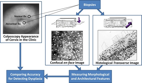
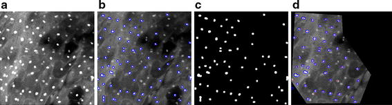
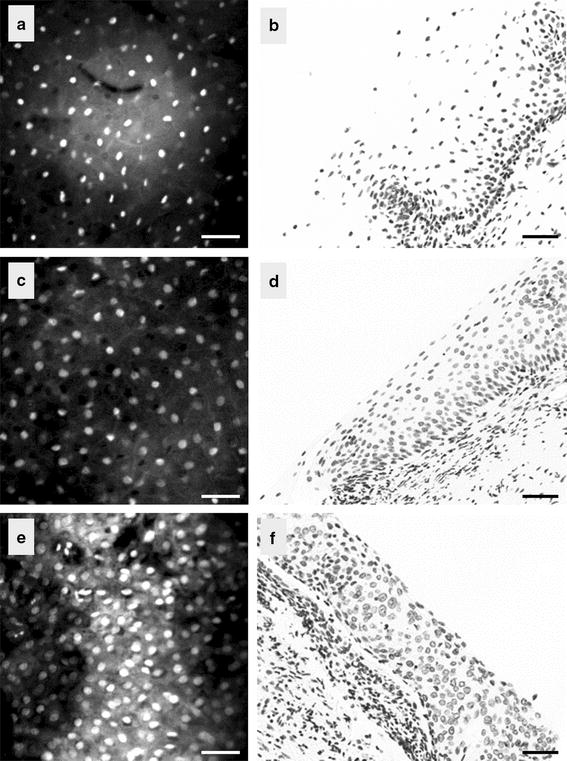
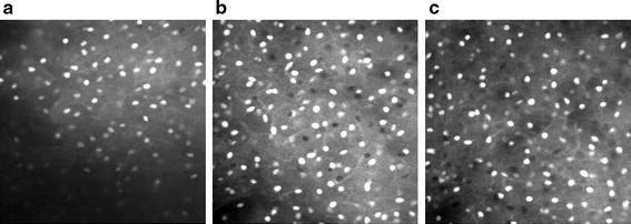
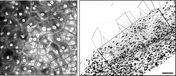
We examined the quantification of ex vivo confocal fluorescence microscopy to differentiate among normal cervical tissue, low-grade Cervical Intraepithelial Neoplasia (CIN), and high-grade CIN. We sought to (1) quantify nuclear morphology and tissue architecture features by analyzing images of cervical biopsies; and (2) determine the accuracy of high-grade CIN detection via confocal microscopy relative to the accuracy of detection by colposcopic impression. Forty-six biopsies obtained from colposcopically normal and abnormal cervical sites were evaluated. Confocal images were acquired at different depths from the epithelial surface and histological images were analyzed using in-house software.
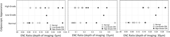
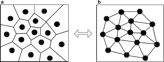
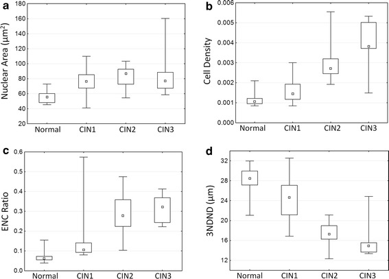
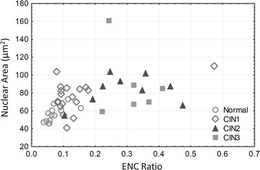
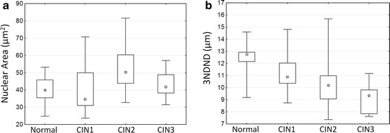
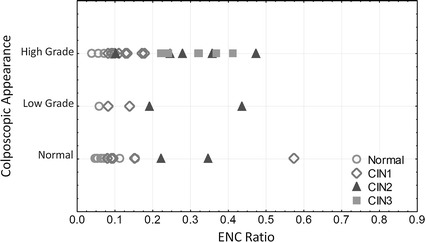
The features calculated from the confocal images compared well with those features obtained from the histological images and histopathological reviews of the specimens (obtained by a gynecologic pathologist). The correlations between two of these features (the nuclear-cytoplasmic ratio and the average of three nearest Delaunay-neighbors distance) and the grade of dysplasia were higher than that of colposcopic impression. The sensitivity of detecting high-grade dysplasia by analysing images collected at the surface of the epithelium, and at 15 and 30 μm below the epithelial surface were respectively 100, 100, and 92 %.
Quantitative analysis of confocal fluorescence images showed its capacity for discriminating high-grade CIN lesions vs. low-grade CIN lesions and normal tissues, at different depth of imaging. This approach could be used to help clinicians identify high-grade CIN in clinical settings.
宫颈癌仍然是一个重大的健康问题,尤其是在发展中国家。阴道镜检查用于检测有异常巴氏涂片病史患者的高级别病变。需要新技术来提高该技术的敏感性和特异性。我们提议测试荧光共聚焦显微镜识别高级别病变的潜力。
我们研究了离体共聚焦荧光显微镜定量分析以区分正常宫颈组织、低级别宫颈上皮内瘤变(CIN)和高级别CIN。我们试图:(1)通过分析宫颈活检图像来量化细胞核形态和组织结构特征;(2)确定共聚焦显微镜检测高级别CIN相对于阴道镜印象检测的准确性。对从阴道镜检查正常和异常宫颈部位获取的46份活检样本进行了评估。从上皮表面不同深度获取共聚焦图像,并使用内部软件分析组织学图像。
从共聚焦图像计算得出的特征与从组织学图像和标本的组织病理学评估(由妇科病理学家获得)得出的特征比较吻合。其中两个特征(核质比和三个最近德劳内邻居距离的平均值)与发育异常分级之间的相关性高于阴道镜印象。通过分析在上皮表面、上皮表面下方15μm和30μm处采集的图像检测高级别发育异常的敏感性分别为100%、100%和92%。
共聚焦荧光图像的定量分析显示了其在不同成像深度区分高级别CIN病变与低级别CIN病变和正常组织的能力。这种方法可用于帮助临床医生在临床环境中识别高级别CIN。