Jentzen Walter, Richter Marion, Nagarajah James, Poeppel Thorsten Dirk, Brandau Wolfgang, Dawes Colin, Bockisch Andreas, Binse Ina
Klinik für Nuklearmedizin, Universität Duisburg-Essen, Hufelandstrasse 55, D-45122, Essen, Germany.
Department of Oral Biology, Faculty of Dentistry, University of Manitoba, 780 Bannatyne Avenue, Winnipeg, Manitoba,, R3E 0W2, Canada.
EJNMMI Phys. 2014 Dec;1(1):100. doi: 10.1186/s40658-014-0100-1. Epub 2014 Dec 6.
The goal of this prospective study was to estimate the absorbed (radiation) doses to salivary glands in radioiodine therapy of thyroid cancer under chewing-gum stimulation using (124)I positron emission tomography (PET)/computed tomography (CT) imaging.
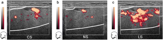
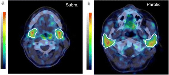
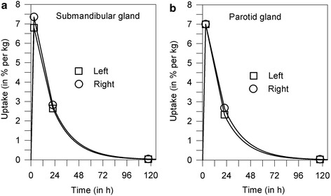
Duplex ultrasonography was conducted in three test persons for visual comparison of the glandular blood flow with three different stimulation types (no stimulation, chewing tasteless gum base, sucking on lemon slices). Ten patients with newly diagnosed differentiated thyroid cancer received (124)I PET/CT dosimetry after thyroidectomy and prior to radioiodine therapy. Patients underwent a series of three (124)I PET/CT scans (4, 24, and ≥96 h after administration of 23 MBq (124)I). They were instructed to chew gum base (tasteless) approximately 20 min after ingesting the (124)I-containing capsule in the course of the first day. Absorbed doses per administered (131)I activity to the salivary glands were calculated and compared with the previously published results of the lemon-juice stimulation and non-stimulation groups.
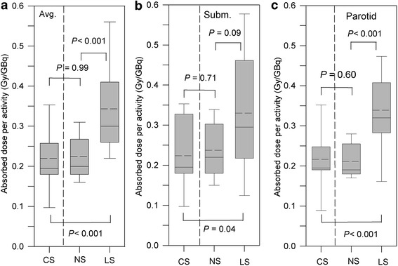
The sonograms in the three test persons showed that glandular blood perfusion by lemon-juice stimulation was clearly increased compared with non-stimulation or chewing of gum base. The sonogram comparison between the chewing-gum stimulation and non-stimulation demonstrated a minor increase of blood flow for the gum base-stimulated salivary glands. The mean ± standard deviation of the absorbed dose per activity under chewing-gum stimulation for the submandibular and parotid glands (within parentheses) was 0.22 ± 0.09 Gy/GBq (0.22 ± 0.08 Gy/GBq). Compared with the absorbed doses of the non-stimulation group, 0.24 ± 0.08 Gy/GBq (0.21 ± 0.05 Gy/GBq), those of the chewing-gum stimulation group showed no significant change (P > 0.60), but the absorbed doses of the lemon-juice stimulation group, 0.35 ± 0.14 Gy/GBq (0.33 ± 0.09 Gy/GBq), were significantly higher (P < 0.04) than those of the chewing-gum stimulation group.
The results suggest that salivary flow induced by chewing gum base does not cause a significant reduction of the salivary gland absorbed dose compared with that in the non-stimulation group. The increased blood flow appears to be a decisive factor causing the increased (131)I absorbed doses in the lemon-juice stimulation group.
本前瞻性研究的目的是利用(124)I 正电子发射断层扫描(PET)/计算机断层扫描(CT)成像,估算在口香糖刺激下甲状腺癌放射性碘治疗中唾液腺的吸收(辐射)剂量。
对三名受试者进行双功超声检查,以视觉比较三种不同刺激类型(无刺激、咀嚼无味口香糖基、吮吸柠檬片)下的腺体血流情况。十名新诊断为分化型甲状腺癌的患者在甲状腺切除术后及放射性碘治疗前接受了(124)I PET/CT 剂量测定。患者在给予 23 MBq(124)I 后进行了一系列三次(124)I PET/CT 扫描(给药后 4、24 和≥96 小时)。在第一天服用含(124)I 胶囊后约 20 分钟,指导他们咀嚼口香糖基(无味)。计算每给予的(131)I 活度对唾液腺的吸收剂量,并与先前发表的柠檬汁刺激组和无刺激组的结果进行比较。
三名受试者的超声图显示,与无刺激或咀嚼口香糖基相比,柠檬汁刺激下的腺体血流灌注明显增加。口香糖刺激组与无刺激组的超声图比较显示,口香糖基刺激的唾液腺血流略有增加。在口香糖刺激下,下颌下腺和腮腺(括号内)每活度吸收剂量的平均值±标准差为 0.22±0.09 Gy/GBq(0.22±0.08 Gy/GBq)。与无刺激组的吸收剂量 0.24±0.08 Gy/GBq(0.21±0.05 Gy/GBq)相比,口香糖刺激组的吸收剂量无显著变化(P>0.60),但柠檬汁刺激组的吸收剂量 0.35±0.14 Gy/GBq(0.33±0.09 Gy/GBq)显著高于口香糖刺激组(P<0.04)。
结果表明,与无刺激组相比,咀嚼口香糖基引起的唾液流动不会导致唾液腺吸收剂量显著降低。血流增加似乎是导致柠檬汁刺激组(131)I 吸收剂量增加的决定性因素。