• 文献检索
  • 文档翻译
  • 深度研究
  • 学术资讯
  • Suppr Zotero 插件Zotero 插件
  • 邀请有礼
  • 套餐&价格
  • 历史记录
应用&插件
Suppr Zotero 插件Zotero 插件浏览器插件Mac 客户端Windows 客户端微信小程序
定价
高级版会员购买积分包购买API积分包
服务
文献检索文档翻译深度研究API 文档MCP 服务
关于我们
关于 Suppr公司介绍联系我们用户协议隐私条款
关注我们

Suppr 超能文献

核心技术专利:CN118964589B侵权必究
粤ICP备2023148730 号-1Suppr @ 2026

文献检索

告别复杂PubMed语法,用中文像聊天一样搜索,搜遍4000万医学文献。AI智能推荐,让科研检索更轻松。

立即免费搜索

文件翻译

保留排版,准确专业,支持PDF/Word/PPT等文件格式,支持 12+语言互译。

免费翻译文档

深度研究

AI帮你快速写综述,25分钟生成高质量综述,智能提取关键信息,辅助科研写作。

立即免费体验

一种使用高密度互补金属氧化物半导体微电极阵列对仓鼠视网膜神经节细胞进行电生理特性分析的方法。

A method for electrophysiological characterization of hamster retinal ganglion cells using a high-density CMOS microelectrode array.

作者信息

Jones Ian L, Russell Thomas L, Farrow Karl, Fiscella Michele, Franke Felix, Müller Jan, Jäckel David, Hierlemann Andreas

机构信息

Bio Engineering Laboratory, Department of Biosystems Science and Engineering, ETH Zurich Basel, Switzerland.

Visual Circuits Laboratory, Neuroelectronics Research Flanders Leuven, Belgium ; NERF, Imec Leuven, Belgium ; Department of Biology, KU Leuven Leuven, Belgium.

出版信息

Front Neurosci. 2015 Oct 13;9:360. doi: 10.3389/fnins.2015.00360. eCollection 2015.

DOI:10.3389/fnins.2015.00360
PMID:26528115
原文链接:https://pmc.ncbi.nlm.nih.gov/articles/PMC4602149/
Abstract

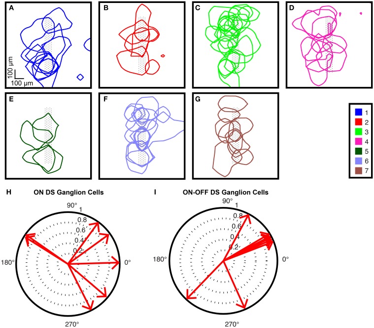
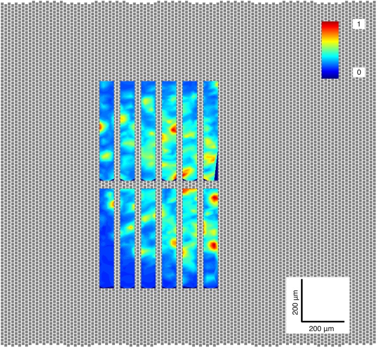
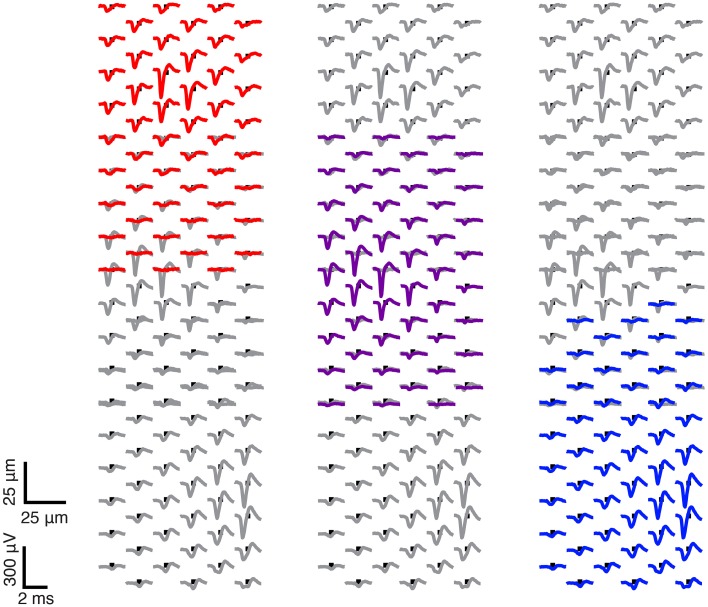
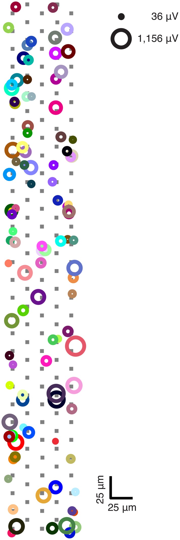
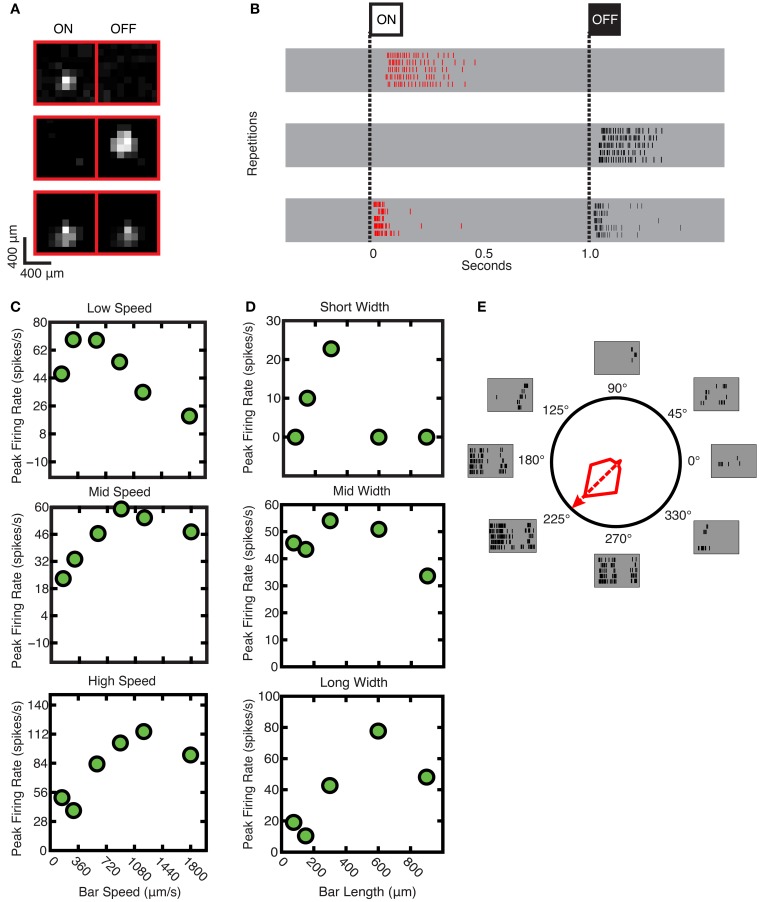
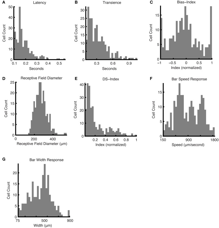
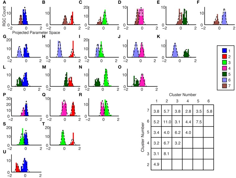
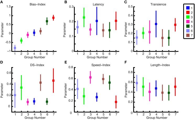
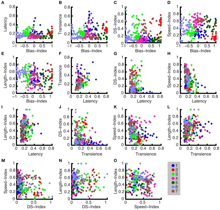
Knowledge of neuronal cell types in the mammalian retina is important for the understanding of human retinal disease and the advancement of sight-restoring technology, such as retinal prosthetic devices. A somewhat less utilized animal model for retinal research is the hamster, which has a visual system that is characterized by an area centralis and a wide visual field with a broad binocular component. The hamster retina is optimally suited for recording on the microelectrode array (MEA), because it intrinsically lies flat on the MEA surface and yields robust, large-amplitude signals. However, information in the literature about hamster retinal ganglion cell functional types is scarce. The goal of our work is to develop a method featuring a high-density (HD) complementary metal-oxide-semiconductor (CMOS) MEA technology along with a sequence of standardized visual stimuli in order to categorize ganglion cells in isolated Syrian Hamster (Mesocricetus auratus) retina. Since the HD-MEA is capable of recording at a higher spatial resolution than most MEA systems (17.5 μm electrode pitch), we were able to record from a large proportion of RGCs within a selected region. Secondly, we chose our stimuli so that they could be run during the experiment without intervention or computation steps. The visual stimulus set was designed to activate the receptive fields of most ganglion cells in parallel and to incorporate various visual features to which different cell types respond uniquely. Based on the ganglion cell responses, basic cell properties were determined: direction selectivity, speed tuning, width tuning, transience, and latency. These properties were clustered to identify ganglion cell types in the hamster retina. Ultimately, we recorded up to a cell density of 2780 cells/mm(2) at 2 mm (42°) from the optic nerve head. Using five parameters extracted from the responses to visual stimuli, we obtained seven ganglion cell types.

摘要

了解哺乳动物视网膜中的神经元细胞类型对于理解人类视网膜疾病以及推进视力恢复技术(如视网膜假体装置)至关重要。仓鼠是一种在视网膜研究中较少使用的动物模型,其视觉系统的特点是有一个中央凹区域和一个具有广泛双眼成分的广阔视野。仓鼠视网膜非常适合在微电极阵列(MEA)上进行记录,因为它本质上平放在MEA表面并产生强大的大幅度信号。然而,文献中关于仓鼠视网膜神经节细胞功能类型的信息很少。我们工作的目标是开发一种方法,该方法采用高密度(HD)互补金属氧化物半导体(CMOS)MEA技术以及一系列标准化视觉刺激,以便对分离的叙利亚仓鼠(Mesocricetus auratus)视网膜中的神经节细胞进行分类。由于HD-MEA能够以比大多数MEA系统更高的空间分辨率进行记录(电极间距为17.5μm),我们能够在选定区域内从很大比例的视网膜神经节细胞(RGCs)进行记录。其次,我们选择的刺激方式使得它们可以在实验过程中无需干预或计算步骤即可运行。视觉刺激集的设计旨在并行激活大多数神经节细胞的感受野,并纳入不同细胞类型独特响应的各种视觉特征。基于神经节细胞的反应,确定了基本的细胞特性:方向选择性、速度调谐、宽度调谐、瞬态性和潜伏期。这些特性被聚类以识别仓鼠视网膜中的神经节细胞类型。最终,我们在距离视神经乳头2mm(42°)处记录到细胞密度高达2780个细胞/mm²。利用从视觉刺激反应中提取的五个参数,我们获得了七种神经节细胞类型。

https://cdn.ncbi.nlm.nih.gov/pmc/blobs/746b/4602149/1ec50c17b950/fnins-09-00360-g0010.jpg
https://cdn.ncbi.nlm.nih.gov/pmc/blobs/746b/4602149/61ead1424347/fnins-09-00360-g0001.jpg
https://cdn.ncbi.nlm.nih.gov/pmc/blobs/746b/4602149/dbd81624c08c/fnins-09-00360-g0002.jpg
https://cdn.ncbi.nlm.nih.gov/pmc/blobs/746b/4602149/7fd32561af29/fnins-09-00360-g0003.jpg
https://cdn.ncbi.nlm.nih.gov/pmc/blobs/746b/4602149/bca9ede65f94/fnins-09-00360-g0004.jpg
https://cdn.ncbi.nlm.nih.gov/pmc/blobs/746b/4602149/ddf860708ec2/fnins-09-00360-g0005.jpg
https://cdn.ncbi.nlm.nih.gov/pmc/blobs/746b/4602149/c74cd47e0bd4/fnins-09-00360-g0006.jpg
https://cdn.ncbi.nlm.nih.gov/pmc/blobs/746b/4602149/2e3e61e6c466/fnins-09-00360-g0007.jpg
https://cdn.ncbi.nlm.nih.gov/pmc/blobs/746b/4602149/1930737acd1b/fnins-09-00360-g0008.jpg
https://cdn.ncbi.nlm.nih.gov/pmc/blobs/746b/4602149/f3055bdc69af/fnins-09-00360-g0009.jpg
https://cdn.ncbi.nlm.nih.gov/pmc/blobs/746b/4602149/1ec50c17b950/fnins-09-00360-g0010.jpg

相似文献

1
A method for electrophysiological characterization of hamster retinal ganglion cells using a high-density CMOS microelectrode array.一种使用高密度互补金属氧化物半导体微电极阵列对仓鼠视网膜神经节细胞进行电生理特性分析的方法。
Front Neurosci. 2015 Oct 13;9:360. doi: 10.3389/fnins.2015.00360. eCollection 2015.
2
Recording from defined populations of retinal ganglion cells using a high-density CMOS-integrated microelectrode array with real-time switchable electrode selection.使用高密度 CMOS 集成微电极阵列对特定人群的视网膜神经节细胞进行记录,该微电极阵列具有实时可切换的电极选择功能。
J Neurosci Methods. 2012 Oct 15;211(1):103-13. doi: 10.1016/j.jneumeth.2012.08.017. Epub 2012 Aug 23.
3
Three Small-Receptive-Field Ganglion Cells in the Mouse Retina Are Distinctly Tuned to Size, Speed, and Object Motion.小鼠视网膜中的三种小感受野神经节细胞对大小、速度和物体运动具有明显的调谐特性。
J Neurosci. 2017 Jan 18;37(3):610-625. doi: 10.1523/JNEUROSCI.2804-16.2016.
4
Reconstruction of natural images from responses of primate retinal ganglion cells.从灵长类视网膜神经节细胞的反应中重建自然图像。
Elife. 2020 Nov 4;9:e58516. doi: 10.7554/eLife.58516.
5
Electrical Imaging of Light-Induced Signals Across and Within Retinal Layers.光诱导信号在视网膜各层之间及层内的电成像。
Front Neurosci. 2020 Nov 19;14:563964. doi: 10.3389/fnins.2020.563964. eCollection 2020.
6
Ananalysis of electrically evoked responses of midget and parasol retinal ganglion cells in different retinal regions.不同视网膜区域中小双极和伞状视网膜神经节细胞电诱发反应的分析。
J Neural Eng. 2022 Mar 30;19(2). doi: 10.1088/1741-2552/ac5b18.
7
Spatiotemporal characteristics of retinal response to network-mediated photovoltaic stimulation.视网膜对网络介导的光电压刺激反应的时空特征
J Neurophysiol. 2018 Feb 1;119(2):389-400. doi: 10.1152/jn.00872.2016. Epub 2017 Oct 18.
8
Investigation of the Functional Retinal Output Using Microelectrode Arrays.使用微电极阵列对视网膜功能输出的研究。
Methods Mol Biol. 2018;1695:81-88. doi: 10.1007/978-1-4939-7407-8_8.
9
Optimization of stimulation parameters for epi-retinal implant based on biosafety consideration.基于生物安全性考虑的视网膜上植体刺激参数的优化。
PLoS One. 2020 Jul 22;15(7):e0236176. doi: 10.1371/journal.pone.0236176. eCollection 2020.
10
High spatial resolution artificial vision inferred from the spiking output of retinal ganglion cells stimulated by optogenetic and electrical means.通过光遗传学和电学手段刺激视网膜神经节细胞的脉冲输出推断出的高空间分辨率人工视觉。
Front Cell Neurosci. 2022 Dec 9;16:1033738. doi: 10.3389/fncel.2022.1033738. eCollection 2022.

引用本文的文献

1
Gap junctions fine-tune ganglion cell signals to equalize response kinetics within a given electrically coupled array.缝隙连接对神经节细胞信号进行微调,以使给定电耦合阵列内的反应动力学达到均衡。
iScience. 2024 May 24;27(6):110099. doi: 10.1016/j.isci.2024.110099. eCollection 2024 Jun 21.
2
Effect of epiretinal electrical stimulation on the glial cells in a rabbit retinal eyecup model.视网膜前电刺激对兔视网膜眼杯模型中神经胶质细胞的影响。
Front Neurosci. 2024 Jan 22;18:1290829. doi: 10.3389/fnins.2024.1290829. eCollection 2024.
3
Transience of the Retinal Output Is Determined by a Great Variety of Circuit Elements.

本文引用的文献

1
High-resolution CMOS MEA platform to study neurons at subcellular, cellular, and network levels.用于在亚细胞、细胞和网络水平研究神经元的高分辨率互补金属氧化物半导体微电极阵列平台。
Lab Chip. 2015 Jul 7;15(13):2767-80. doi: 10.1039/c5lc00133a. Epub 2015 May 14.
2
The types of retinal ganglion cells: current status and implications for neuronal classification.视网膜神经节细胞的类型:现状及对神经元分类的影响。
Annu Rev Neurosci. 2015 Jul 8;38:221-46. doi: 10.1146/annurev-neuro-071714-034120. Epub 2015 Apr 9.
3
Bayes optimal template matching for spike sorting - combining fisher discriminant analysis with optimal filtering.
视网膜输出的瞬态由各种各样的电路元件决定。
Cells. 2022 Feb 25;11(5):810. doi: 10.3390/cells11050810.
4
Technologies to Study Action Potential Propagation With a Focus on HD-MEAs.聚焦于高密度微电极阵列研究动作电位传播的技术。
Front Cell Neurosci. 2019 Apr 26;13:159. doi: 10.3389/fncel.2019.00159. eCollection 2019.
5
Non-parametric Physiological Classification of Retinal Ganglion Cells in the Mouse Retina.小鼠视网膜中视网膜神经节细胞的非参数生理分类
Front Cell Neurosci. 2018 Dec 7;12:481. doi: 10.3389/fncel.2018.00481. eCollection 2018.
6
A method for single-neuron chronic recording from the retina in awake mice.一种在清醒小鼠的视网膜上进行单神经元慢性记录的方法。
Science. 2018 Jun 29;360(6396):1447-1451. doi: 10.1126/science.aas9160.
7
Technical feasibility study for production of tailored multielectrode arrays and patterning of arranged neuronal networks.定制多电极阵列生产及有序神经网络图案化的技术可行性研究。
PLoS One. 2018 Feb 23;13(2):e0192647. doi: 10.1371/journal.pone.0192647. eCollection 2018.
8
Mechanisms creating transient and sustained photoresponses in mammalian retinal ganglion cells.在哺乳动物视网膜神经节细胞中产生瞬态和持续光反应的机制。
J Gen Physiol. 2017 Mar 6;149(3):335-353. doi: 10.1085/jgp.201611720. Epub 2017 Feb 2.
用于尖峰分类的贝叶斯最优模板匹配——将Fisher判别分析与最优滤波相结合。
J Comput Neurosci. 2015 Jun;38(3):439-59. doi: 10.1007/s10827-015-0547-7. Epub 2015 Feb 5.
4
Revealing neuronal function through microelectrode array recordings.通过微电极阵列记录揭示神经元功能。
Front Neurosci. 2015 Jan 6;8:423. doi: 10.3389/fnins.2014.00423. eCollection 2014.
5
Retinal implants: a systematic review.视网膜植入物:系统评价。
Br J Ophthalmol. 2014 Jul;98(7):852-6. doi: 10.1136/bjophthalmol-2013-303708.
6
High-density microelectrode array recordings and real-time spike sorting for closed-loop experiments: an emerging technology to study neural plasticity.高密度微电极阵列记录和实时尖峰分类用于闭环实验:研究神经可塑性的新兴技术。
Front Neural Circuits. 2012 Dec 20;6:105. doi: 10.3389/fncir.2012.00105. eCollection 2012.
7
Features and functions of nonlinear spatial integration by retinal ganglion cells.视网膜神经节细胞的非线性空间整合的特征与功能。
J Physiol Paris. 2013 Nov;107(5):338-48. doi: 10.1016/j.jphysparis.2012.12.001. Epub 2012 Dec 20.
8
Neural stimulation for visual rehabilitation: advances and challenges.用于视觉康复的神经刺激:进展与挑战。
J Physiol Paris. 2013 Nov;107(5):421-31. doi: 10.1016/j.jphysparis.2012.10.003. Epub 2012 Nov 10.
9
Visual stimulation reverses the directional preference of direction-selective retinal ganglion cells.视觉刺激会改变方向选择性视网膜神经节细胞的方向偏好。
Neuron. 2012 Nov 8;76(3):518-25. doi: 10.1016/j.neuron.2012.08.041.
10
Recording from defined populations of retinal ganglion cells using a high-density CMOS-integrated microelectrode array with real-time switchable electrode selection.使用高密度 CMOS 集成微电极阵列对特定人群的视网膜神经节细胞进行记录,该微电极阵列具有实时可切换的电极选择功能。
J Neurosci Methods. 2012 Oct 15;211(1):103-13. doi: 10.1016/j.jneumeth.2012.08.017. Epub 2012 Aug 23.