Aoyagi Ranko, Hayashi Takaaki, Tsuneoka Hiroshi
Department of Ophthalmology, The Jikei University School of Medicine, Tokyo, Japan.
Int Med Case Rep J. 2015 Nov 11;8:291-4. doi: 10.2147/IMCRJ.S95442. eCollection 2015.
The purpose of this study was to report optical coherence tomography (OCT) and angiographic findings in a patient with pregnancy-induced hypertension (PIH).
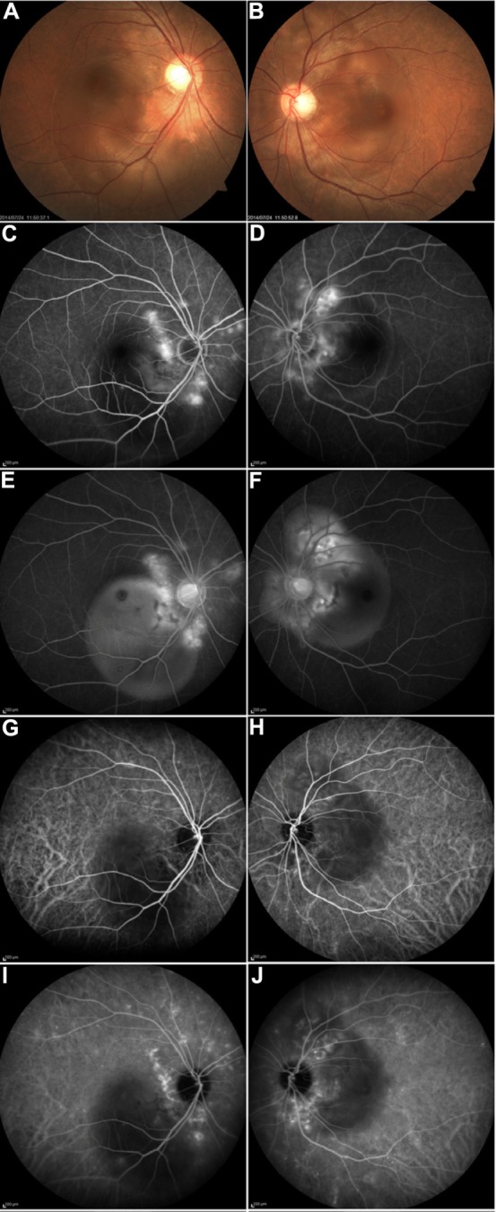
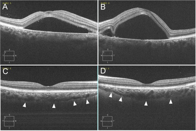
A 39-year-old woman, who was diagnosed with PIH, reported blurred and distorted vision at 5 days after an emergency cesarean delivery. OCT revealed a large serous retinal detachment (SRD) that included areas in the macula, along with an increased choroidal thickness noted in both eyes. Indocyanine green angiograms indicated delayed filling of the choroidal circulation in the early phase but choroidal hyperpermeability in the mid-phase. The SRD was gradually resolving without any treatment except for antihypertensive drugs. At 40 days after the initial examination, OCT revealed both the disappearance of the SRD and marked improvement of the choroidal thickening.
Ophthalmologists need to be aware that PIH can cause choroidal ischemia, a breakdown of the outer blood-retinal barrier, and lead to the development of SRD.
本研究旨在报告一名妊娠高血压综合征(PIH)患者的光学相干断层扫描(OCT)及血管造影结果。
一名39岁被诊断为PIH的女性,在急诊剖宫产术后5天出现视力模糊及视物变形。OCT显示双眼存在累及黄斑区的大片浆液性视网膜脱离(SRD),同时脉络膜厚度增加。吲哚菁绿血管造影显示早期脉络膜循环充盈延迟,但中期脉络膜通透性增加。除降压药物外未进行任何治疗,SRD逐渐消退。初次检查后40天,OCT显示SRD消失且脉络膜增厚明显改善。
眼科医生应意识到PIH可导致脉络膜缺血、外层血视网膜屏障破坏,并引发SRD。