Hayasaka Daisuke, Nishi Kodai, Fuchigami Takeshi, Shiogama Kazuya, Onouchi Takanori, Shimada Satoshi, Tsutsumi Yutaka, Morita Kouichi
Department of Virology, Institute of Tropical Medicine, Nagasaki University, Sakamoto, Nagasaki, Japan.
Leading Graduate School Program, Nagasaki University, Sakamoto, Nagasaki, Japan.
Oncotarget. 2016 Jan 5;7(1):140-7. doi: 10.18632/oncotarget.6645.
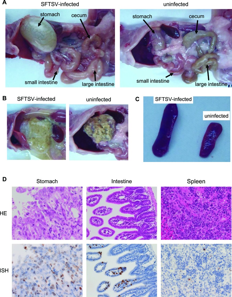
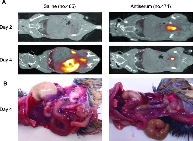
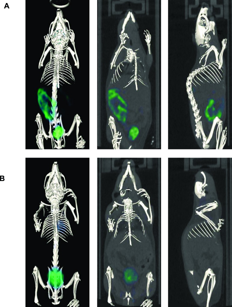
Severe fever with thrombocytopenia syndrome (SFTS) is an emerging disease that causes fever, enteritis, thrombocytopenia, and leucopenia and can be fatal in up to 30% of cases. However, the mechanism of severe disease is not fully understood. Molecular imaging approaches, such as positron-emission tomography (PET), are functional in vivo imaging techniques that provide real-time dynamics of disease progression, assessments of pharmacokinetics, and diagnoses for disease progression. Molecular imaging also potentially provides useful approaches to explore the pathogenesis of viral infections. Thus, the purpose of this study was to image the pathological features of SFTSV infection in vivo by PET imaging. In a mouse model, we showed that 18F-FDG accumulations clearly identified the intestinal tract site as a pathological site. We also demonstrated that 18F-FDG PET imaging can assess disease progression and response to antiserum therapy within the same individual. This is the first report demonstrating a molecular imaging strategy for SFTSV infection. Our results provide potentially useful information for preclinical studies such as the elucidation of the mechanism of SFTSV infection in vivo and the assessment of drugs for SFTS treatment.
严重发热伴血小板减少综合征(SFTS)是一种新出现的疾病,可导致发热、肠炎、血小板减少和白细胞减少,高达30%的病例可能会死亡。然而,重症疾病的机制尚未完全明确。分子成像方法,如正电子发射断层扫描(PET),是功能性的体内成像技术,可提供疾病进展的实时动态、药代动力学评估以及疾病进展的诊断。分子成像还可能为探索病毒感染的发病机制提供有用的方法。因此,本研究的目的是通过PET成像对SFTSV感染的病理特征进行体内成像。在小鼠模型中,我们发现18F-FDG的聚集清晰地将肠道部位确定为病理部位。我们还证明18F-FDG PET成像可以评估同一动物体内的疾病进展和对抗血清治疗的反应。这是第一份展示针对SFTSV感染的分子成像策略的报告。我们的结果为临床前研究提供了潜在有用的信息,如阐明SFTSV体内感染机制以及评估SFTS治疗药物。