• 文献检索
  • 文档翻译
  • 深度研究
  • 学术资讯
  • Suppr Zotero 插件Zotero 插件
  • 邀请有礼
  • 套餐&价格
  • 历史记录
应用&插件
Suppr Zotero 插件Zotero 插件浏览器插件Mac 客户端Windows 客户端微信小程序
定价
高级版会员购买积分包购买API积分包
服务
文献检索文档翻译深度研究API 文档MCP 服务
关于我们
关于 Suppr公司介绍联系我们用户协议隐私条款
关注我们

Suppr 超能文献

核心技术专利:CN118964589B侵权必究
粤ICP备2023148730 号-1Suppr @ 2026

文献检索

告别复杂PubMed语法,用中文像聊天一样搜索,搜遍4000万医学文献。AI智能推荐,让科研检索更轻松。

立即免费搜索

文件翻译

保留排版,准确专业,支持PDF/Word/PPT等文件格式,支持 12+语言互译。

免费翻译文档

深度研究

AI帮你快速写综述,25分钟生成高质量综述,智能提取关键信息,辅助科研写作。

立即免费体验

滤泡性淋巴瘤的分类:计算机辅助对病理学家分级的影响

Classification of follicular lymphoma: the effect of computer aid on pathologists grading.

作者信息

Fauzi Mohammad Faizal Ahmad, Pennell Michael, Sahiner Berkman, Chen Weijie, Shana'ah Arwa, Hemminger Jessica, Gru Alejandro, Kurt Habibe, Losos Michael, Joehlin-Price Amy, Kavran Christina, Smith Stephen M, Nowacki Nicholas, Mansor Sharmeen, Lozanski Gerard, Gurcan Metin N

机构信息

Faculty of Engineering, Multimedia University, 63100, Cyberjaya, Selangor, Malaysia.

Division of Biostatistics, College of Public Health, The Ohio State University, Columbus, OH, USA.

出版信息

BMC Med Inform Decis Mak. 2015 Dec 30;15:115. doi: 10.1186/s12911-015-0235-6.

DOI:10.1186/s12911-015-0235-6
PMID:26715518
原文链接:https://pmc.ncbi.nlm.nih.gov/articles/PMC4696238/
Abstract

BACKGROUND

Follicular lymphoma (FL) is one of the most common lymphoid malignancies in the western world. FL cases are stratified into three histological grades based on the average centroblast count per high power field (HPF). The centroblast count is performed manually by the pathologist using an optical microscope and hematoxylin and eosin (H&E) stained tissue section. Although this is the current clinical practice, it suffers from high inter- and intra-observer variability and is vulnerable to sampling bias.

METHODS

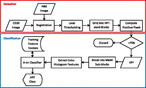
In this paper, we present a system, called Follicular Lymphoma Grading System (FLAGS), to assist the pathologist in grading FL cases. We also assess the effect of FLAGS on accuracy of expert and inexperienced readers. FLAGS automatically identifies possible HPFs for examination by analyzing H&E and CD20 stains, before classifying them into low or high risk categories. The pathologist is first asked to review the slides according to the current routine clinical practice, before being presented with FLAGS classification via color-coded map. The accuracy of the readers with and without FLAGS assistance is measured.

RESULTS

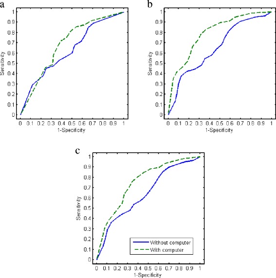
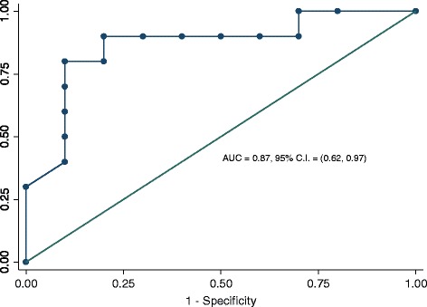
FLAGS was used by four experts (board-certified hematopathologists) and seven pathology residents on 20 FL slides. Access to FLAGS improved overall reader accuracy with the biggest improvement seen among residents. An average AUC value of 0.75 was observed which generally indicates "acceptable" diagnostic performance.

CONCLUSIONS

The results of this study show that FLAGS can be useful in increasing the pathologists' accuracy in grading the tissue. To the best of our knowledge, this study measure, for the first time, the effect of computerized image analysis on pathologists' grading of follicular lymphoma. When fully developed, such systems have the potential to reduce sampling bias by examining an increased proportion of HPFs within follicle regions, as well as to reduce inter- and intra-reader variability.

摘要

背景

滤泡性淋巴瘤(FL)是西方世界最常见的淋巴恶性肿瘤之一。根据每个高倍视野(HPF)中的中心母细胞平均计数,FL病例被分为三个组织学等级。中心母细胞计数由病理学家使用光学显微镜和苏木精-伊红(H&E)染色的组织切片手动进行。尽管这是目前的临床实践,但它存在较高的观察者间和观察者内变异性,并且容易受到抽样偏差的影响。

方法

在本文中,我们提出了一种名为滤泡性淋巴瘤分级系统(FLAGS)的系统,以协助病理学家对FL病例进行分级。我们还评估了FLAGS对专家和经验不足的读者准确性的影响。FLAGS通过分析H&E和CD20染色自动识别可能需要检查的HPF,然后将它们分为低风险或高风险类别。病理学家首先被要求根据当前的常规临床实践审查幻灯片,然后通过颜色编码图呈现FLAGS分类。测量有和没有FLAGS协助的读者的准确性。

结果

四名专家(获得委员会认证的血液病理学家)和七名病理住院医师在2​​0张FL幻灯片上使用了FLAGS。使用FLAGS提高了读者的整体准确性,住院医师的提高最为明显。观察到平均AUC值为0.75,这通常表明诊断性能“可接受”。

结论

本研究结果表明,FLAGS有助于提高病理学家对组织分级的准确性。据我们所知,本研究首次测量了计算机图像分析对病理学家滤泡性淋巴瘤分级的影响。当充分开发时,此类系统有可能通过检查卵泡区域内增加比例的HPF来减少抽样偏差,并减少读者间和读者内的变异性。

https://cdn.ncbi.nlm.nih.gov/pmc/blobs/e847/4696238/8faadb452bb9/12911_2015_235_Fig3_HTML.jpg
https://cdn.ncbi.nlm.nih.gov/pmc/blobs/e847/4696238/7c3e8df96431/12911_2015_235_Fig1_HTML.jpg
https://cdn.ncbi.nlm.nih.gov/pmc/blobs/e847/4696238/ddabd0abcbb1/12911_2015_235_Fig2_HTML.jpg
https://cdn.ncbi.nlm.nih.gov/pmc/blobs/e847/4696238/8faadb452bb9/12911_2015_235_Fig3_HTML.jpg
https://cdn.ncbi.nlm.nih.gov/pmc/blobs/e847/4696238/7c3e8df96431/12911_2015_235_Fig1_HTML.jpg
https://cdn.ncbi.nlm.nih.gov/pmc/blobs/e847/4696238/ddabd0abcbb1/12911_2015_235_Fig2_HTML.jpg
https://cdn.ncbi.nlm.nih.gov/pmc/blobs/e847/4696238/8faadb452bb9/12911_2015_235_Fig3_HTML.jpg

相似文献

1
Classification of follicular lymphoma: the effect of computer aid on pathologists grading.滤泡性淋巴瘤的分类:计算机辅助对病理学家分级的影响
BMC Med Inform Decis Mak. 2015 Dec 30;15:115. doi: 10.1186/s12911-015-0235-6.
2
Computer-aided detection of centroblasts for follicular lymphoma grading using adaptive likelihood-based cell segmentation.基于自适应似然的细胞分割的滤泡性淋巴瘤分级中中心母细胞的计算机辅助检测。
IEEE Trans Biomed Eng. 2010 Oct;57(10):2613-6. doi: 10.1109/TBME.2010.2055058. Epub 2010 Jun 28.
3
A study of criteria for grading follicular lymphoma using a cell type classifier from pathology images based on complementary-label learning.一项基于互补标签学习,使用来自病理图像的细胞类型分类器对滤泡性淋巴瘤进行分级的标准研究。
Micron. 2024 Sep;184:103663. doi: 10.1016/j.micron.2024.103663. Epub 2024 May 30.
4
Computer-assisted quantification of CD3+ T cells in follicular lymphoma.滤泡性淋巴瘤中CD3 + T细胞的计算机辅助定量分析
Cytometry A. 2017 Jun;91(6):609-621. doi: 10.1002/cyto.a.23049. Epub 2017 Jan 22.
5
The Utility of Phosphohistone H3 (PHH3) in Follicular Lymphoma Grading: A Comparative Study With Ki-67 and H&E Mitotic Count.磷酸组蛋白 H3(PHH3)在滤泡性淋巴瘤分级中的应用:与 Ki-67 和 H&E 有丝分裂计数的比较研究。
Am J Clin Pathol. 2019 May 3;151(6):542-550. doi: 10.1093/ajcp/aqz003.
6
Inter-reader variability in follicular lymphoma grading: Conventional and digital reading.滤泡性淋巴瘤分级中的阅片者间变异性:传统阅片与数字阅片
J Pathol Inform. 2013 Oct 29;4:30. doi: 10.4103/2153-3539.120747. eCollection 2013.
7
Utility of SKP2 and MIB-1 in grading follicular lymphoma using quantitative imaging analysis.利用定量成像分析评估SKP2和MIB-1在滤泡性淋巴瘤分级中的作用。
Hum Pathol. 2007 Jun;38(6):878-82. doi: 10.1016/j.humpath.2006.11.021. Epub 2007 Mar 12.
8
Detection of follicles from IHC-stained slides of follicular lymphoma using iterative watershed.使用迭代分水岭算法从滤泡性淋巴瘤的免疫组织化学染色切片中检测滤泡。
IEEE Trans Biomed Eng. 2010 Oct;57(10):2609-12. doi: 10.1109/TBME.2010.2058111. Epub 2010 Jul 15.
9
Grading of follicular lymphoma in cytological samples.细胞学样本中滤泡性淋巴瘤的分级
Cytopathology. 2016 Dec;27(6):390-397. doi: 10.1111/cyt.12319. Epub 2016 Feb 12.
10
Informatics Approaches to Address New Challenges in the Classification of Lymphoid Malignancies.应对淋巴恶性肿瘤分类新挑战的信息学方法。
JCO Clin Cancer Inform. 2018;2. doi: 10.1200/CCI.17.00039. Epub 2018 Feb 9.

引用本文的文献

1
Mitigating bias in prostate cancer diagnosis using synthetic data for improved AI driven Gleason grading.利用合成数据减轻前列腺癌诊断中的偏差以改进人工智能驱动的 Gleason 分级。
NPJ Precis Oncol. 2025 May 23;9(1):151. doi: 10.1038/s41698-025-00934-5.
2
Role of artificial intelligence in haematolymphoid diagnostics.人工智能在血液淋巴系统诊断中的作用。
Histopathology. 2025 Jan;86(1):58-68. doi: 10.1111/his.15327. Epub 2024 Oct 22.
3
Automated scoring methods for quantitative interpretation of Tumour infiltrating lymphocytes (TILs) in breast cancer: a systematic review.

本文引用的文献

1
A multi-resolution textural approach to diagnostic neuropathology reporting.一种用于诊断神经病理学报告的多分辨率纹理方法。
J Neurooncol. 2015 Sep;124(3):393-402. doi: 10.1007/s11060-015-1872-4. Epub 2015 Aug 9.
2
Multireader multicase reader studies with binary agreement data: simulation, analysis, validation, and sizing.具有二元一致性数据的多读者多病例读者研究:模拟、分析、验证和规模确定。
J Med Imaging (Bellingham). 2014 Oct;1(3):031011. doi: 10.1117/1.JMI.1.3.031011. Epub 2014 Dec 4.
3
Heterogeneity assessment of histological tissue sections in whole slide images.
用于乳腺癌肿瘤浸润淋巴细胞(TILs)定量解读的自动化评分方法:系统综述。
BMC Cancer. 2024 Sep 30;24(1):1202. doi: 10.1186/s12885-024-12962-8.
4
Detection of centroblast cells in H&E stained whole slide image based on object detection.基于目标检测在苏木精-伊红(H&E)染色的全玻片图像中检测中心母细胞。
Front Med (Lausanne). 2024 Feb 7;11:1303982. doi: 10.3389/fmed.2024.1303982. eCollection 2024.
5
Comparison of Ki67 Proliferation Index in Gastrointestinal Non-Hodgkin Large B-Cell Lymphomas: The Conventional Method of Evaluation or AI Evaluation?胃肠道非霍奇金大B细胞淋巴瘤中Ki67增殖指数的比较:传统评估方法还是人工智能评估?
Diagnostics (Basel). 2023 Aug 27;13(17):2775. doi: 10.3390/diagnostics13172775.
6
Creating learning health systems and the emerging role of biomedical informatics.创建学习型健康系统及生物医学信息学的新作用。
Learn Health Syst. 2021 Mar 11;6(1):e10259. doi: 10.1002/lrh2.10259. eCollection 2022 Jan.
7
Machine Learning Applications in the Diagnosis of Benign and Malignant Hematological Diseases.机器学习在良性和恶性血液疾病诊断中的应用
Clin Hematol Int. 2020 Dec 21;3(1):13-20. doi: 10.2991/chi.k.201130.001. eCollection 2021 Mar.
8
Report on computational assessment of Tumor Infiltrating Lymphocytes from the International Immuno-Oncology Biomarker Working Group.国际免疫肿瘤生物标志物工作组关于肿瘤浸润淋巴细胞的计算评估报告。
NPJ Breast Cancer. 2020 May 12;6:16. doi: 10.1038/s41523-020-0154-2. eCollection 2020.
9
Artificial Intelligence and Digital Microscopy Applications in Diagnostic Hematopathology.人工智能与数字显微镜在血液病理学诊断中的应用
Cancers (Basel). 2020 Mar 26;12(4):797. doi: 10.3390/cancers12040797.
10
Informatics Approaches to Address New Challenges in the Classification of Lymphoid Malignancies.应对淋巴恶性肿瘤分类新挑战的信息学方法。
JCO Clin Cancer Inform. 2018;2. doi: 10.1200/CCI.17.00039. Epub 2018 Feb 9.
全切片图像中组织切片的异质性评估。
Comput Med Imaging Graph. 2015 Jun;42:51-5. doi: 10.1016/j.compmedimag.2014.11.006. Epub 2014 Nov 20.
4
The average receiver operating characteristic curve in multireader multicase imaging studies.多阅片者多病例影像研究中的平均受试者工作特征曲线。
Br J Radiol. 2014 Aug;87(1040):20140016. doi: 10.1259/bjr.20140016. Epub 2014 Jun 2.
5
Inter-reader variability in follicular lymphoma grading: Conventional and digital reading.滤泡性淋巴瘤分级中的阅片者间变异性:传统阅片与数字阅片
J Pathol Inform. 2013 Oct 29;4:30. doi: 10.4103/2153-3539.120747. eCollection 2013.
6
Histopathological image analysis for centroblasts classification through dimensionality reduction approaches.通过降维方法对中心母细胞进行组织病理学图像分析分类。
Cytometry A. 2014 Mar;85(3):242-55. doi: 10.1002/cyto.a.22432. Epub 2013 Dec 26.
7
An Efficient Computational Framework for the Analysis of Whole Slide Images: Application to Follicular Lymphoma Immunohistochemistry.一种用于全切片图像分析的高效计算框架:在滤泡性淋巴瘤免疫组织化学中的应用。
J Comput Sci. 2012 Sep 1;3(5):269-279. doi: 10.1016/j.jocs.2012.01.009. Epub 2012 Mar 6.
8
A general framework for the segmentation of follicular lymphoma virtual slides.滤泡性淋巴瘤虚拟切片分割的通用框架。
Comput Med Imaging Graph. 2012 Sep;36(6):442-51. doi: 10.1016/j.compmedimag.2012.05.003. Epub 2012 Jun 18.
9
Automatic detection of follicular regions in H&E images using iterative shape index.使用迭代形状指数自动检测 H&E 图像中的滤泡区域。
Comput Med Imaging Graph. 2011 Oct-Dec;35(7-8):592-602. doi: 10.1016/j.compmedimag.2011.03.001. Epub 2011 Apr 20.
10
Detection of follicles from IHC-stained slides of follicular lymphoma using iterative watershed.使用迭代分水岭算法从滤泡性淋巴瘤的免疫组织化学染色切片中检测滤泡。
IEEE Trans Biomed Eng. 2010 Oct;57(10):2609-12. doi: 10.1109/TBME.2010.2058111. Epub 2010 Jul 15.