Stavely Rhian, Robinson Ainsley M, Miller Sarah, Boyd Richard, Sakkal Samy, Nurgali Kulmira
Centre for Chronic Disease, College of Health and Biomedicine, Western Centre for Health, Research and Education, Sunshine Hospital, 176 Furlong road, Melbourne, 3021, Victoria, Australia.
Department of Anatomy and Developmental Biology, Monash University, 19 Innovation Walk, Clayton, 3800, Victoria, Australia.
Stem Cell Res Ther. 2015 Dec 30;6:263. doi: 10.1186/s13287-015-0254-3.
The use of mesenchymal stem cells (MSCs) to treat inflammatory bowel disease (IBD) is of great interest because of their immunomodulatory properties. Damage to the enteric nervous system (ENS) is implicated in IBD pathophysiology and disease progression. The most commonly used model to study inflammation-induced changes to the ENS is 2,4,6-trinitrobenzene-sulfonate acid (TNBS)-induced colitis in guinea pigs; however, no studies using guinea pig MSCs in colitis have been performed. This study aims to isolate and characterise guinea pig MSCs and then test their therapeutic potential for the treatment of enteric neuropathy associated with intestinal inflammation.
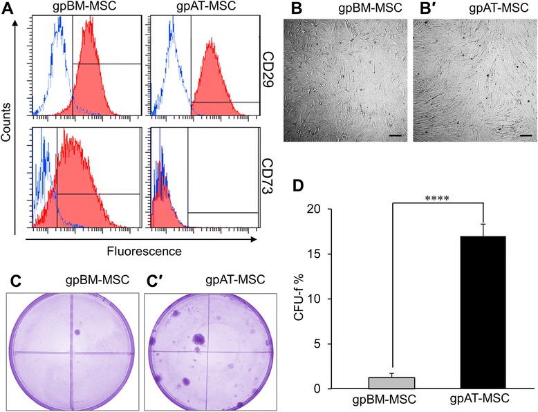
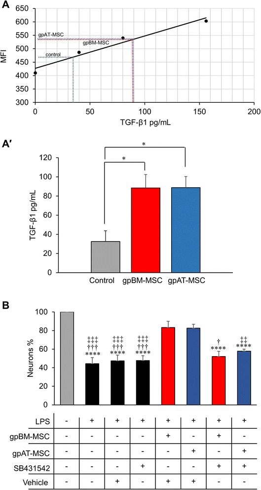
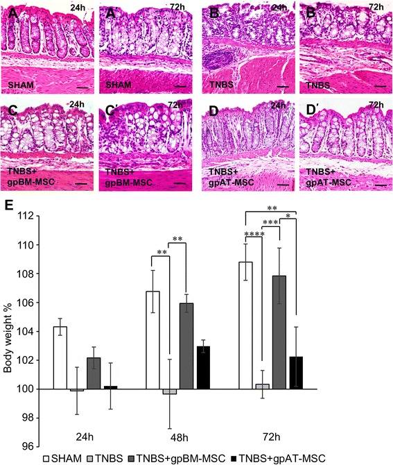
MSCs from guinea pig bone marrow and adipose tissue were isolated and characterised in vitro. In in vivo experiments, guinea pigs received either TNBS for the induction of colitis or sham treatment by enema. MSCs were administered at a dose of 1 × 10(6) cells via enema 3 h after the induction of colitis. Colon tissues were collected 24 and 72 h after TNBS administration to assess the level of inflammation and damage to the ENS. The secretion of transforming growth factor-β1 (TGF-β1) was analysed in MSC conditioned medium by flow cytometry.
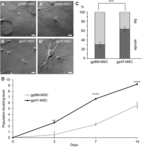
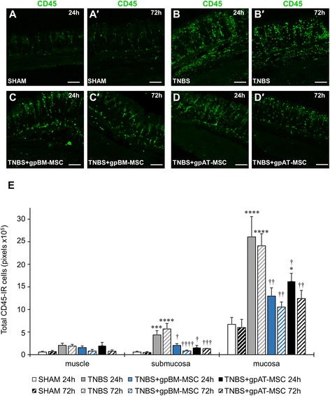
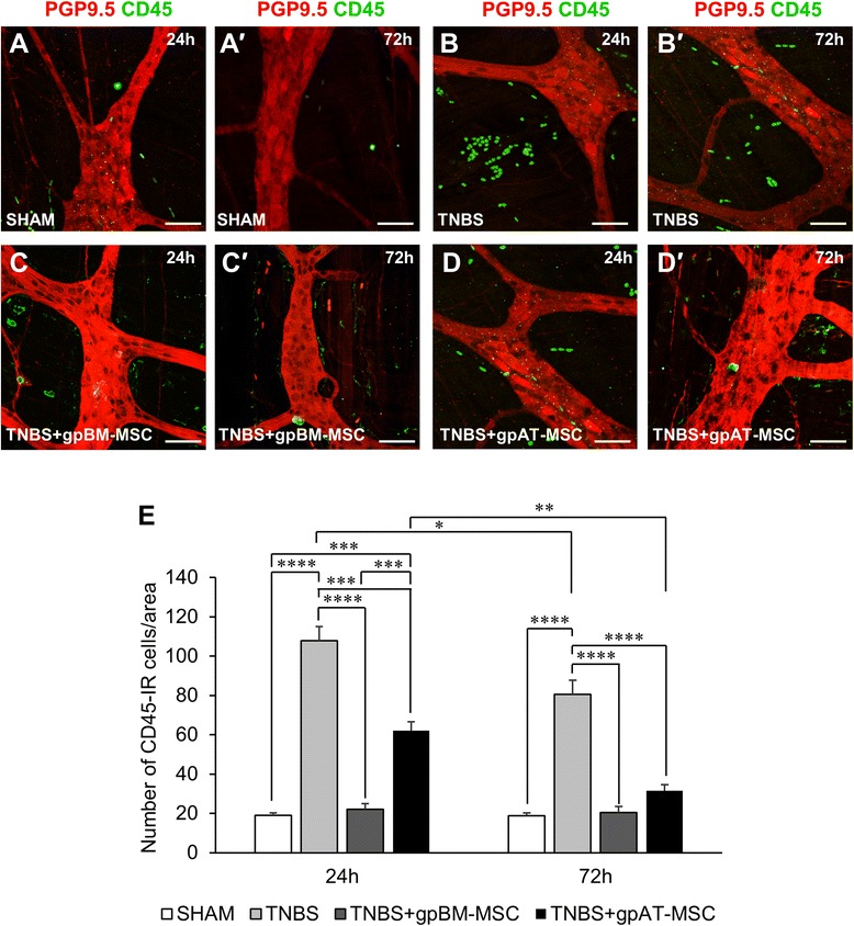
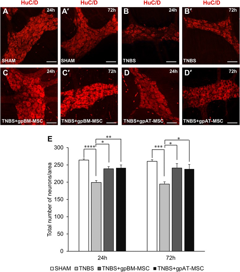
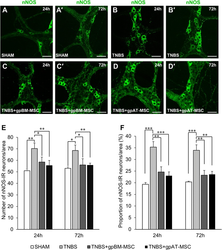
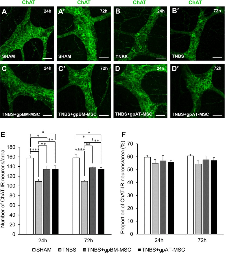
Cells isolated from both sources were adherent to plastic, multipotent and expressed some human MSC surface markers. In vitro characterisation revealed distinct differences in growth kinetics, clonogenicity and cell morphology between MSC types. In an in vivo model of TNBS-induced colitis, guinea pig bone marrow MSCs were comparatively more efficacious than adipose tissue MSCs in attenuating weight loss, colonic tissue damage and leukocyte infiltration into the mucosa and myenteric plexus. MSCs from both sources were equally neuroprotective in the amelioration of enteric neuronal loss and changes to the neurochemical coding of neuronal subpopulations. MSCs from both sources secreted TGF-β1 which exerted neuroprotective effects in vitro.
This study is the first evaluating the functional capacity of guinea pig bone marrow and adipose tissue-derived MSCs and providing evidence of their neuroprotective value in an animal model of colitis. In vitro characteristics of MSCs cannot be extrapolated to their therapeutic efficacy. TGF-β1 released by both types of MSCs might have contributed to the attenuation of enteric neuropathy associated with colitis.
由于间充质干细胞(MSC)具有免疫调节特性,其在治疗炎症性肠病(IBD)方面备受关注。肠神经系统(ENS)损伤与IBD的病理生理学及疾病进展有关。研究炎症诱导的ENS变化最常用的模型是2,4,6 -三硝基苯磺酸(TNBS)诱导的豚鼠结肠炎;然而,尚未有使用豚鼠MSC治疗结肠炎的研究。本研究旨在分离并鉴定豚鼠MSC,然后测试其治疗与肠道炎症相关的肠神经病变的潜力。
从豚鼠骨髓和脂肪组织中分离MSC并进行体外鉴定。在体内实验中,豚鼠接受TNBS灌肠诱导结肠炎或假处理。在诱导结肠炎3小时后,通过灌肠给予剂量为1×10⁶个细胞的MSC。在给予TNBS后24小时和72小时收集结肠组织,以评估炎症水平和ENS损伤情况。通过流式细胞术分析MSC条件培养基中转化生长因子-β1(TGF-β1)的分泌情况。
从两种来源分离的细胞均贴壁生长、具有多能性并表达一些人MSC表面标志物。体外鉴定显示不同类型的MSC在生长动力学、克隆形成能力和细胞形态方面存在明显差异。在TNBS诱导的结肠炎体内模型中,豚鼠骨髓MSC在减轻体重减轻、结肠组织损伤以及白细胞浸润至黏膜和肌间神经丛方面比脂肪组织MSC更有效。两种来源的MSC在改善肠神经元丢失和神经元亚群神经化学编码变化方面具有同等的神经保护作用。两种来源的MSC均分泌TGF-β1,其在体外发挥神经保护作用。
本研究首次评估了豚鼠骨髓和脂肪组织来源的MSC的功能能力,并在结肠炎动物模型中提供了其神经保护价值的证据。MSC的体外特性不能外推至其治疗效果。两种类型的MSC释放的TGF-β1可能有助于减轻与结肠炎相关的肠神经病变。