Erber W N, McLachlan J
Haematology Department, Royal North Shore Hospital, St Leonards, New South Wales, Australia.
J Clin Pathol. 1989 Nov;42(11):1201-5. doi: 10.1136/jcp.42.11.1201.
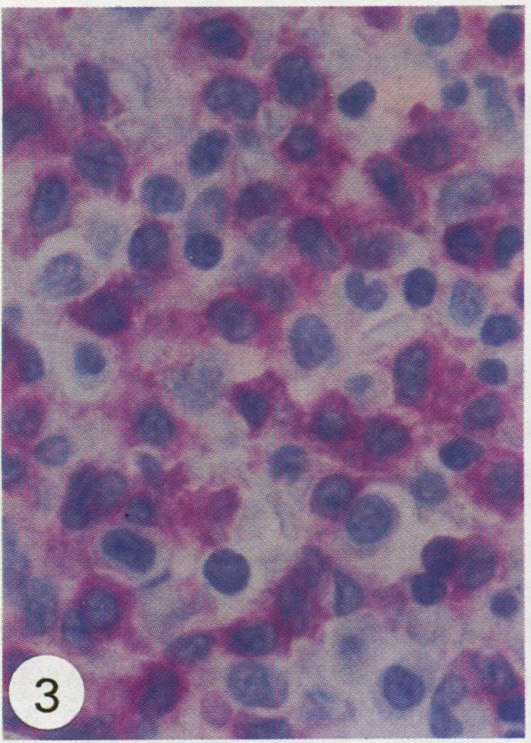
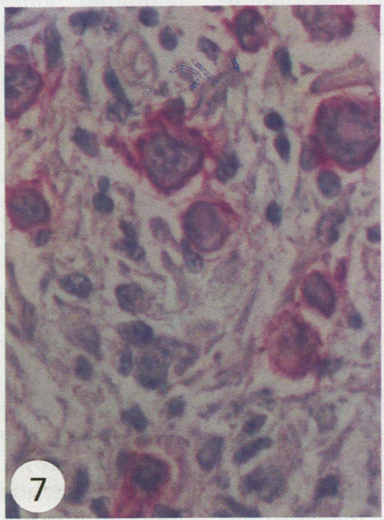
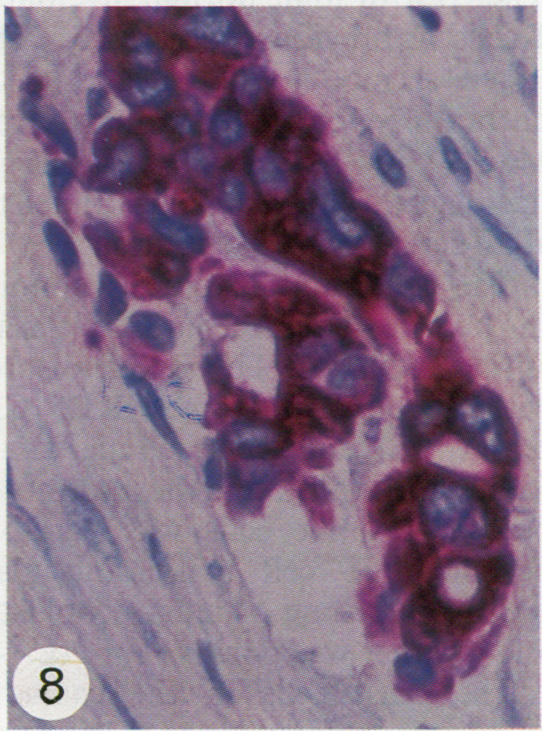
The immunoalkaline phosphatase (APAAP) technique was applied to the labelling of decalcified sections of formalin fixed, paraffin wax embedded bone marrow trephine biopsy specimens. A panel of monoclonal antibodies reactive with haemopoietic and epithelial antigens, which survive routine formalin fixation, was assessed on 72 cases of haematological malignancy (including acute and chronic leukaemias and lymphomas) showing bone marrow infiltration. The APAAP method showed clear distinct labelling of antigen positive cells without loss of antigens due to decalcification. Both normal or reactive single cells present in the sample and neoplastic cell populations could be identified morphologically and their antigenic phenotype and cellular origin, whether lymphoid or myeloid, established. The application of the APAAP method to routinely prepared paraffin wax embedded trephines has many advantages over the assessment of specially prepared cryostat sections of bone marrow.
免疫碱性磷酸酶(APAAP)技术应用于福尔马林固定、石蜡包埋的骨髓环钻活检标本脱钙切片的标记。在72例显示骨髓浸润的血液系统恶性肿瘤(包括急性和慢性白血病及淋巴瘤)中,评估了一组与经常规福尔马林固定后仍存活的造血和上皮抗原反应的单克隆抗体。APAAP方法显示抗原阳性细胞标记清晰、明显,且未因脱钙而导致抗原丢失。样本中存在的正常或反应性单个细胞以及肿瘤细胞群均可通过形态学鉴定,并确定其抗原表型和细胞来源,无论是淋巴样还是髓样。与评估专门制备的骨髓低温切片相比,将APAAP方法应用于常规制备的石蜡包埋环钻切片有许多优点。