• 文献检索
  • 文档翻译
  • 深度研究
  • 学术资讯
  • Suppr Zotero 插件Zotero 插件
  • 邀请有礼
  • 套餐&价格
  • 历史记录
应用&插件
Suppr Zotero 插件Zotero 插件浏览器插件Mac 客户端Windows 客户端微信小程序
定价
高级版会员购买积分包购买API积分包
服务
文献检索文档翻译深度研究API 文档MCP 服务
关于我们
关于 Suppr公司介绍联系我们用户协议隐私条款
关注我们

Suppr 超能文献

核心技术专利:CN118964589B侵权必究
粤ICP备2023148730 号-1Suppr @ 2026

文献检索

告别复杂PubMed语法,用中文像聊天一样搜索,搜遍4000万医学文献。AI智能推荐,让科研检索更轻松。

立即免费搜索

文件翻译

保留排版,准确专业,支持PDF/Word/PPT等文件格式,支持 12+语言互译。

免费翻译文档

深度研究

AI帮你快速写综述,25分钟生成高质量综述,智能提取关键信息,辅助科研写作。

立即免费体验

钛植入物上的喷丸处理:粗糙度、成骨细胞功能及与骨水泥结合的影响

Peen treatment on a titanium implant: effect of roughness, osteoblast cell functions, and bonding with bone cement.

作者信息

Khandaker Morshed, Riahinezhad Shahram, Sultana Fariha, Vaughan Melville B, Knight Joshua, Morris Tracy L

机构信息

Department of Engineering & Physics, University of Central Oklahoma, Edmond, OK, USA; Center for Interdisciplinary Biomedical Education and Research, University of Central Oklahoma, Edmond, OK, USA.

Department of Engineering & Physics, University of Central Oklahoma, Edmond, OK, USA.

出版信息

Int J Nanomedicine. 2016 Feb 4;11:585-94. doi: 10.2147/IJN.S89376. eCollection 2016.

DOI:10.2147/IJN.S89376
PMID:26893563
原文链接:https://pmc.ncbi.nlm.nih.gov/articles/PMC4745826/
Abstract

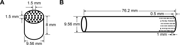
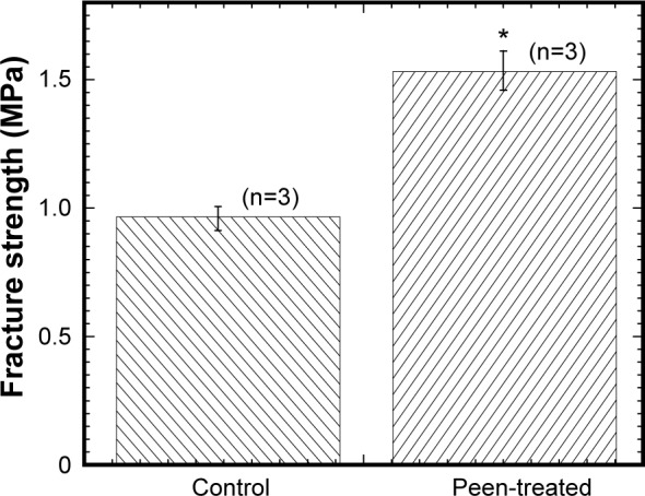
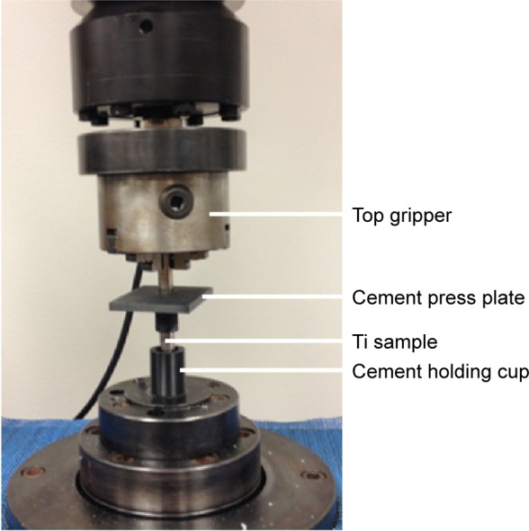
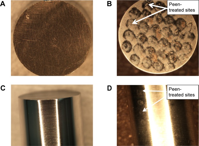
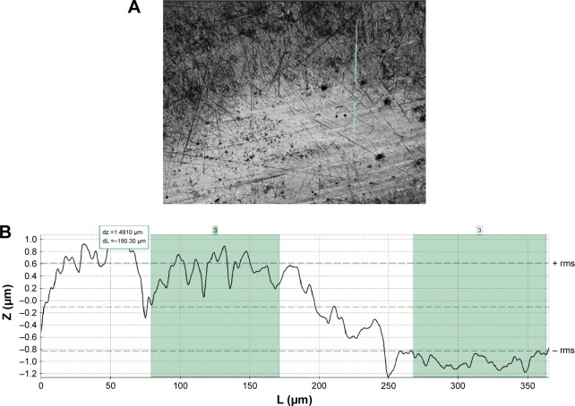
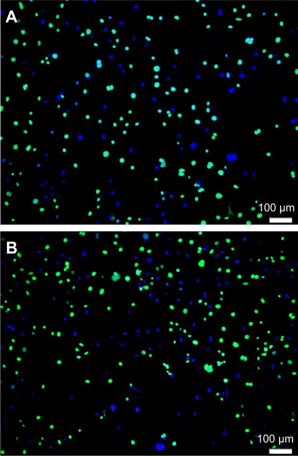
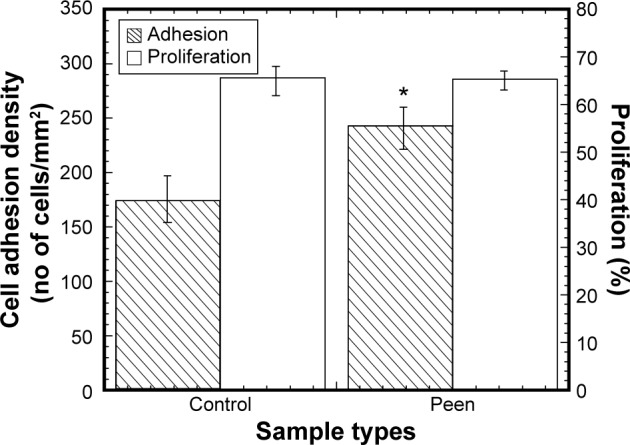
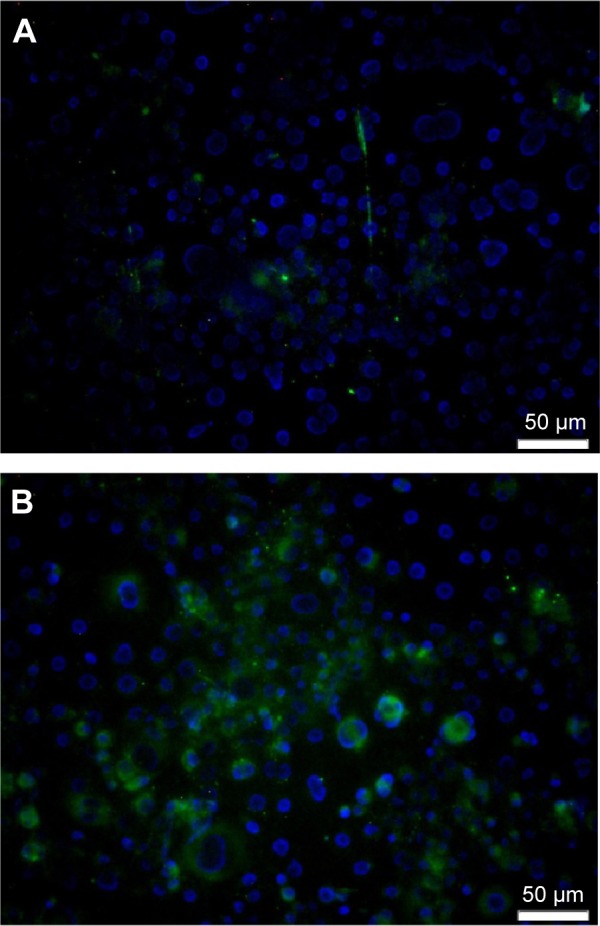
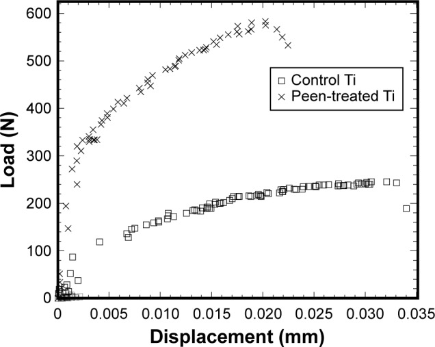
Implant failure due to poor integration of the implant with the surrounding biomaterial is a common problem in various orthopedic and orthodontic surgeries. Implant fixation mostly depends upon the implant surface topography. Micron to nanosize circular-shaped groove architecture with adequate surface roughness can enhance the mechanical interlock and osseointegration of an implant with the host tissue and solve its poor fixation problem. Such groove architecture can be created on a titanium (Ti) alloy implant by laser peening treatment. Laser peening produces deep, residual compressive stresses in the surfaces of metal parts, delivering increased fatigue life and damage tolerance. The scientific novelty of this study is the controlled deposition of circular-shaped rough spot groove using laser peening technique and understanding the effect of the treatment techniques for improving the implant surface properties. The hypothesis of this study was that implant surface grooves created by controlled laser peen treatment can improve the mechanical and biological responses of the implant with the adjoining biomaterial. The objective of this study was to measure how the controlled laser-peened groove architecture on Ti influences its osteoblast cell functions and bonding strength with bone cement. This study determined the surface roughness and morphology of the peen-treated Ti. In addition, this study compared the osteoblast cell functions (adhesion, proliferation, and differentiation) between control and peen-treated Ti samples. Finally, this study measured the fracture strength between each kind of Ti samples and bone cement under static loading. This study found that laser peen treatment on Ti significantly changed the surface architecture of the Ti, which led to enhanced osteoblast cell adhesion and differentiation on Ti implants and fracture strength of Ti-bone cement interfaces compared with values of untreated Ti samples. Therefore, the laser peen treatment method has the potential to improve the biomechanical functions of Ti implants.

摘要

由于植入物与周围生物材料的整合不佳而导致的植入失败是各种骨科和正畸手术中常见的问题。植入物的固定主要取决于植入物的表面形貌。具有适当表面粗糙度的微米到纳米尺寸的圆形凹槽结构可以增强植入物与宿主组织的机械互锁和骨整合,并解决其固定不佳的问题。这种凹槽结构可以通过激光喷丸处理在钛(Ti)合金植入物上形成。激光喷丸在金属部件表面产生深的残余压应力,从而提高疲劳寿命和损伤容限。本研究的科学新颖之处在于使用激光喷丸技术可控地沉积圆形粗糙点凹槽,并了解处理技术对改善植入物表面性能的影响。本研究的假设是,通过可控激光喷丸处理创建的植入物表面凹槽可以改善植入物与相邻生物材料的机械和生物学反应。本研究的目的是测量Ti上可控激光喷丸凹槽结构如何影响其成骨细胞功能以及与骨水泥的结合强度。本研究确定了喷丸处理后Ti的表面粗糙度和形貌。此外,本研究比较了对照和喷丸处理的Ti样品之间的成骨细胞功能(粘附、增殖和分化)。最后,本研究测量了每种Ti样品与骨水泥在静态加载下的断裂强度。本研究发现,对Ti进行激光喷丸处理显著改变了Ti的表面结构,与未处理的Ti样品相比,这导致Ti植入物上的成骨细胞粘附和分化增强,以及Ti-骨水泥界面的断裂强度提高。因此,激光喷丸处理方法有可能改善Ti植入物的生物力学功能。

https://cdn.ncbi.nlm.nih.gov/pmc/blobs/6bc1/4745826/9bbc0d1362e1/ijn-11-585Fig12.jpg
https://cdn.ncbi.nlm.nih.gov/pmc/blobs/6bc1/4745826/b713aaa1aa2d/ijn-11-585Fig1.jpg
https://cdn.ncbi.nlm.nih.gov/pmc/blobs/6bc1/4745826/73d57a39bbed/ijn-11-585Fig2.jpg
https://cdn.ncbi.nlm.nih.gov/pmc/blobs/6bc1/4745826/84330d4faa13/ijn-11-585Fig3.jpg
https://cdn.ncbi.nlm.nih.gov/pmc/blobs/6bc1/4745826/15c804822ddb/ijn-11-585Fig4.jpg
https://cdn.ncbi.nlm.nih.gov/pmc/blobs/6bc1/4745826/cb7fa5e04969/ijn-11-585Fig5.jpg
https://cdn.ncbi.nlm.nih.gov/pmc/blobs/6bc1/4745826/300908341c31/ijn-11-585Fig6.jpg
https://cdn.ncbi.nlm.nih.gov/pmc/blobs/6bc1/4745826/231498e65fce/ijn-11-585Fig7.jpg
https://cdn.ncbi.nlm.nih.gov/pmc/blobs/6bc1/4745826/9f3ab242877c/ijn-11-585Fig8.jpg
https://cdn.ncbi.nlm.nih.gov/pmc/blobs/6bc1/4745826/3512a236ac90/ijn-11-585Fig9.jpg
https://cdn.ncbi.nlm.nih.gov/pmc/blobs/6bc1/4745826/2db96913479c/ijn-11-585Fig10.jpg
https://cdn.ncbi.nlm.nih.gov/pmc/blobs/6bc1/4745826/17bdd1db63cd/ijn-11-585Fig11.jpg
https://cdn.ncbi.nlm.nih.gov/pmc/blobs/6bc1/4745826/9bbc0d1362e1/ijn-11-585Fig12.jpg

相似文献

1
Peen treatment on a titanium implant: effect of roughness, osteoblast cell functions, and bonding with bone cement.钛植入物上的喷丸处理:粗糙度、成骨细胞功能及与骨水泥结合的影响
Int J Nanomedicine. 2016 Feb 4;11:585-94. doi: 10.2147/IJN.S89376. eCollection 2016.
2
Plasma nitriding of titanium alloy: Effect of roughness, hardness, biocompatibility, and bonding with bone cement.钛合金的等离子体氮化:粗糙度、硬度、生物相容性以及与骨水泥结合的影响。
Biomed Mater Eng. 2016 Nov 25;27(5):461-474. doi: 10.3233/BME-161600.
3
Fracture toughness of titanium-cement interfaces: effects of fibers and loading angles.钛-水泥界面的断裂韧性:纤维和加载角度的影响。
Int J Nanomedicine. 2014 Apr 1;9:1689-97. doi: 10.2147/IJN.S59253. eCollection 2014.
4
In vitro fibroblast and pre-osteoblastic cellular responses on laser surface modified Ti-6Al-4V.激光表面改性Ti-6Al-4V上的体外成纤维细胞和前成骨细胞反应
Biomed Mater. 2014 Dec 29;10(1):015007. doi: 10.1088/1748-6041/10/1/015007.
5
Spark anodization of titanium-zirconium alloy: surface characterization and bioactivity assessment.钛锆合金的火花阳极氧化:表面表征与生物活性评估。
J Mater Sci Mater Med. 2015 Aug;26(8):221. doi: 10.1007/s10856-015-5555-7. Epub 2015 Aug 11.
6
Response of MG63 osteoblast-like cells to titanium and titanium alloy is dependent on surface roughness and composition.MG63 成骨样细胞对钛及钛合金的反应取决于表面粗糙度和成分。
Biomaterials. 1998 Dec;19(23):2219-32. doi: 10.1016/s0142-9612(98)00144-6.
7
Osseointegration improvement by plasma electrolytic oxidation of modified titanium alloys surfaces.通过对改性钛合金表面进行等离子体电解氧化改善骨整合
J Mater Sci Mater Med. 2015 Feb;26(2):72. doi: 10.1007/s10856-015-5408-4. Epub 2015 Jan 29.
8
Innovative surface modification of Ti-6Al-4V alloy with a positive effect on osteoblast proliferation and fatigue performance.对Ti-6Al-4V合金进行创新表面改性,对成骨细胞增殖和疲劳性能有积极影响。
Mater Sci Eng C Mater Biol Appl. 2014 Jun 1;39:371-9. doi: 10.1016/j.msec.2014.03.024. Epub 2014 Mar 17.
9
Arachidonic acid and prostaglandin E2 influence human osteoblast (MG63) response to titanium surface roughness.花生四烯酸和前列腺素E2影响人成骨细胞(MG63)对钛表面粗糙度的反应。
J Oral Implantol. 2008;34(6):303-12. doi: 10.1563/1548-1336-34.6.303.
10
The role of titanium implant surface modification with hydroxyapatite nanoparticles in progressive early bone-implant fixation in vivo.羟基磷灰石纳米颗粒改性钛种植体表面在体内渐进性早期骨-种植体固定中的作用。
Int J Oral Maxillofac Implants. 2009 Sep-Oct;24(5):808-16.

引用本文的文献

1
Unraveling Residual Stress Distribution Characteristics of 6061-T6 Aluminum Alloy Induced by Laser Shock Peening.揭示激光冲击强化诱导6061-T6铝合金残余应力分布特征
Materials (Basel). 2024 Jul 14;17(14):3484. doi: 10.3390/ma17143484.
2
LASER as a tool for surface modification of dental biomaterials: A review.激光作为牙科生物材料表面改性的工具:综述
Heliyon. 2023 Jun 20;9(6):e17457. doi: 10.1016/j.heliyon.2023.e17457. eCollection 2023 Jun.
3
Human Gingival Fibroblast and Osteoblast Behavior on Groove-Milled Zirconia Implant Surfaces.

本文引用的文献

1
The effect of spark plasma sintering on lithium disilicate glass-ceramics.火花等离子烧结对锂硅玻璃陶瓷的影响。
Dent Mater. 2015 Oct;31(10):e226-35. doi: 10.1016/j.dental.2015.07.001. Epub 2015 Jul 23.
2
Innovative surface modification of Ti-6Al-4V alloy with a positive effect on osteoblast proliferation and fatigue performance.对Ti-6Al-4V合金进行创新表面改性,对成骨细胞增殖和疲劳性能有积极影响。
Mater Sci Eng C Mater Biol Appl. 2014 Jun 1;39:371-9. doi: 10.1016/j.msec.2014.03.024. Epub 2014 Mar 17.
3
A new bioassay identifies proliferation ratios of fibroblasts and myofibroblasts.
人牙龈成纤维细胞和成骨细胞在沟槽铣削氧化锆种植体表面的行为。
Materials (Basel). 2022 Mar 28;15(7):2481. doi: 10.3390/ma15072481.
4
Evaluation of Polycaprolactone Electrospun Nanofiber-Composites for Artificial Skin Based on Dermal Fibroblast Culture.基于真皮成纤维细胞培养的聚己内酯电纺纳米纤维复合材料用于人工皮肤的评估。
Bioengineering (Basel). 2022 Jan 6;9(1):19. doi: 10.3390/bioengineering9010019.
5
Laser-Induced Microgrooves Improve the Mechanical Responses of Cemented Implant Systems.激光诱导微槽改善骨水泥植入系统的力学响应。
Micromachines (Basel). 2020 Apr 29;11(5):466. doi: 10.3390/mi11050466.
6
The Effect of Surface Modification of Ti13Zr13Nb Alloy on Adhesion of Antibiotic and Nanosilver-Loaded Bone Cement Coatings Dedicated for Application as Spacers.Ti13Zr13Nb合金表面改性对用作间隔物的载抗生素和纳米银骨水泥涂层附着力的影响。
Materials (Basel). 2019 Sep 12;12(18):2964. doi: 10.3390/ma12182964.
7
miR-338-3p regulates osteoclastogenesis via targeting IKKβ gene.微小RNA-338-3p通过靶向IKKβ基因调控破骨细胞生成。
In Vitro Cell Dev Biol Anim. 2019 Apr;55(4):243-251. doi: 10.1007/s11626-019-00325-8. Epub 2019 Mar 18.
8
Effect of Collagen-Polycaprolactone Nanofibers Matrix Coating on the Cytocompatibility and Bone Responses of Titanium.胶原蛋白-聚己内酯纳米纤维基质涂层对钛的细胞相容性和骨反应的影响
J Med Biol Eng. 2018 Apr;38(2):197-210. doi: 10.1007/s40846-017-0312-7. Epub 2017 Jul 24.
9
Fortifying the Bone-Implant Interface Part 1: An In Vitro Evaluation of 3D-Printed and TPS Porous Surfaces.强化骨-种植体界面 第1部分:3D打印和TPS多孔表面的体外评估
Int J Spine Surg. 2017 Jun 1;11(3):15. doi: 10.14444/4015. eCollection 2017.
10
Microgroove and Collagen-poly(ε-caprolactone) Nanofiber Mesh Coating Improves the Mechanical Stability and Osseointegration of Titanium Implants.微槽与胶原-聚(ε-己内酯)纳米纤维网涂层可改善钛种植体的机械稳定性和骨整合。
Nanomaterials (Basel). 2017 Jun 13;7(6):145. doi: 10.3390/nano7060145.
一种新的生物测定法可鉴定成纤维细胞和平滑肌成纤维细胞的增殖率。
Cell Biol Int. 2014 Aug;38(8):981-6. doi: 10.1002/cbin.10289. Epub 2014 May 6.
4
Fracture toughness of titanium-cement interfaces: effects of fibers and loading angles.钛-水泥界面的断裂韧性:纤维和加载角度的影响。
Int J Nanomedicine. 2014 Apr 1;9:1689-97. doi: 10.2147/IJN.S59253. eCollection 2014.
5
Comparison of Phenotypic Characterization between Differentiated Osteoblasts from Stem Cells and Calvaria Osteoblasts In vitro.干细胞来源的分化成骨细胞与颅骨成骨细胞体外表型特征的比较
Int J Prev Med. 2013 Feb;4(2):180-6.
6
Micro and nano MgO particles for the improvement of fracture toughness of bone-cement interfaces.用于提高骨水泥界面断裂韧性的微纳级氧化镁颗粒。
J Biomech. 2013 Mar 15;46(5):1035-9. doi: 10.1016/j.jbiomech.2012.12.006. Epub 2013 Jan 16.
7
Assessment of compromised fracture healing.骨折愈合不良的评估。
J Am Acad Orthop Surg. 2012 May;20(5):273-82. doi: 10.5435/JAAOS-20-05-273.
8
Generation and differentiation of microtissues from multipotent precursor cells for use in tissue engineering.多能前体细胞生成和分化微组织用于组织工程。
Nat Protoc. 2011 Oct 13;6(11):1726-35. doi: 10.1038/nprot.2011.394.
9
The size effect of crystalline inclusions on the fracture modes in glass-ceramic materials.微晶夹杂对玻璃陶瓷材料断裂模式的尺寸效应。
J Phys Condens Matter. 2007 Jul 4;19(26):266209. doi: 10.1088/0953-8984/19/26/266209. Epub 2007 Jun 7.
10
Bone remodelling at a glance.骨重塑概览。
J Cell Sci. 2011 Apr 1;124(Pt 7):991-8. doi: 10.1242/jcs.063032.