• 文献检索
  • 文档翻译
  • 深度研究
  • 学术资讯
  • Suppr Zotero 插件Zotero 插件
  • 邀请有礼
  • 套餐&价格
  • 历史记录
应用&插件
Suppr Zotero 插件Zotero 插件浏览器插件Mac 客户端Windows 客户端微信小程序
定价
高级版会员购买积分包购买API积分包
服务
文献检索文档翻译深度研究API 文档MCP 服务
关于我们
关于 Suppr公司介绍联系我们用户协议隐私条款
关注我们

Suppr 超能文献

核心技术专利:CN118964589B侵权必究
粤ICP备2023148730 号-1Suppr @ 2026

文献检索

告别复杂PubMed语法,用中文像聊天一样搜索,搜遍4000万医学文献。AI智能推荐,让科研检索更轻松。

立即免费搜索

文件翻译

保留排版,准确专业,支持PDF/Word/PPT等文件格式,支持 12+语言互译。

免费翻译文档

深度研究

AI帮你快速写综述,25分钟生成高质量综述,智能提取关键信息,辅助科研写作。

立即免费体验

血管生成芽过程中内皮细胞与细胞外基质之间机械相互作用的三维表征

Three-Dimensional Characterization of Mechanical Interactions between Endothelial Cells and Extracellular Matrix during Angiogenic Sprouting.

作者信息

Du Yue, Herath Sahan C B, Wang Qing-guo, Wang Dong-an, Asada H Harry, Chen Peter C Y

机构信息

Department of Mechanical Engineering, National University of Singapore, Singapore.

BioSystems and Micromechanics Interdisciplinary Research Group, Singapore-MIT Alliance for Research and Technology Program, Singapore.

出版信息

Sci Rep. 2016 Feb 23;6:21362. doi: 10.1038/srep21362.

DOI:10.1038/srep21362
PMID:26903154
原文链接:https://pmc.ncbi.nlm.nih.gov/articles/PMC4763258/
Abstract

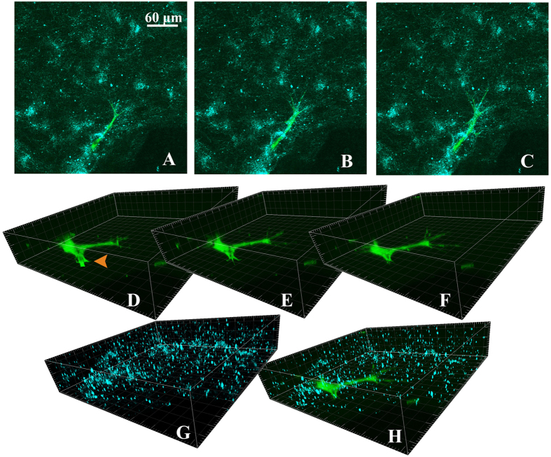
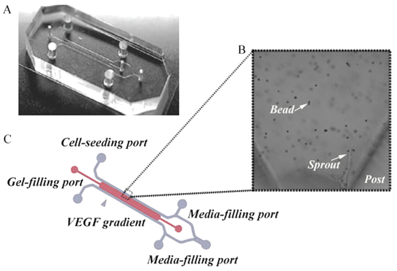
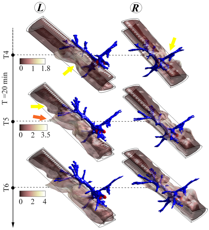
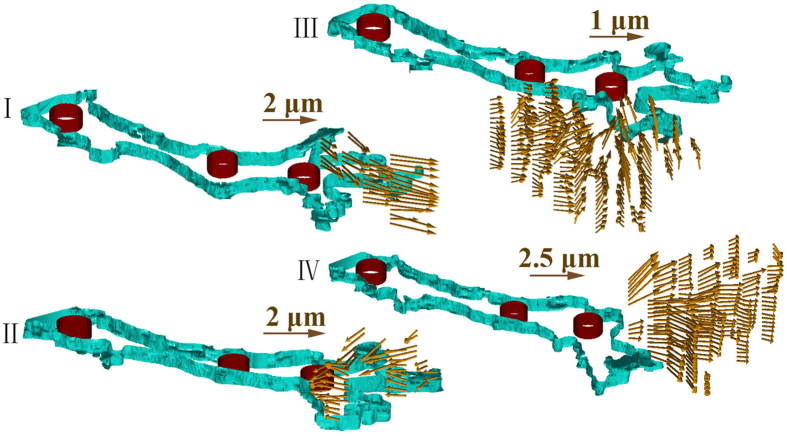
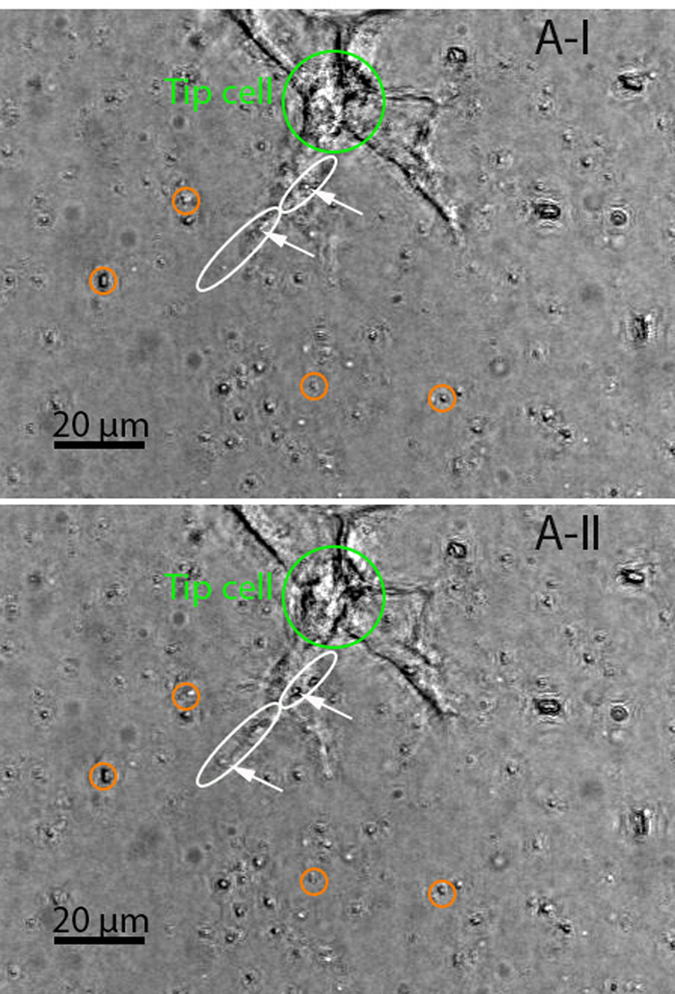
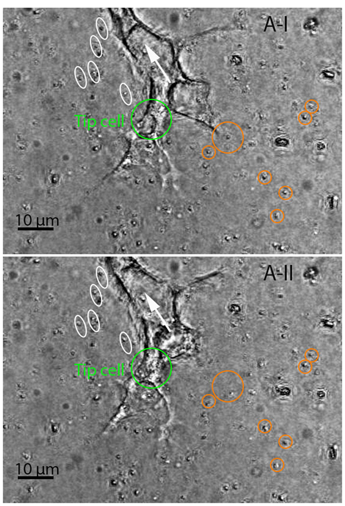
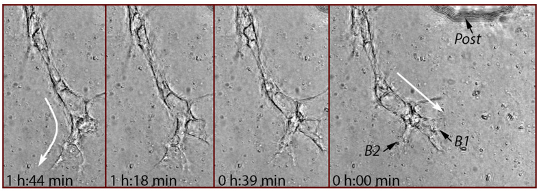
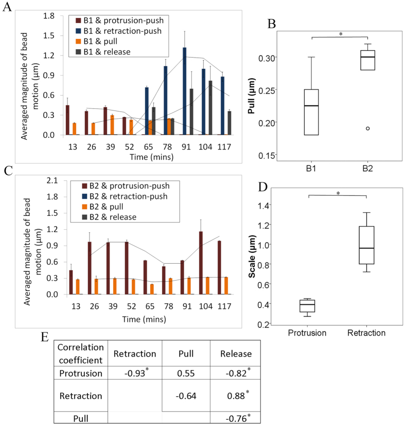
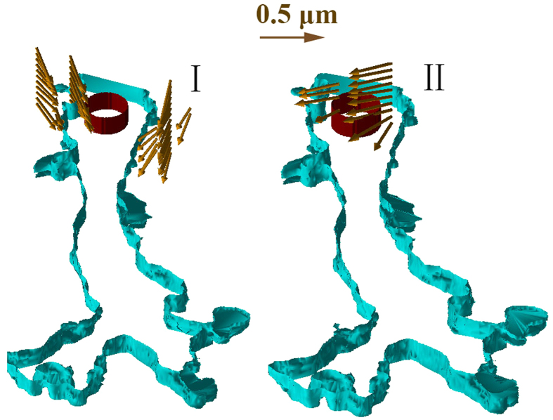
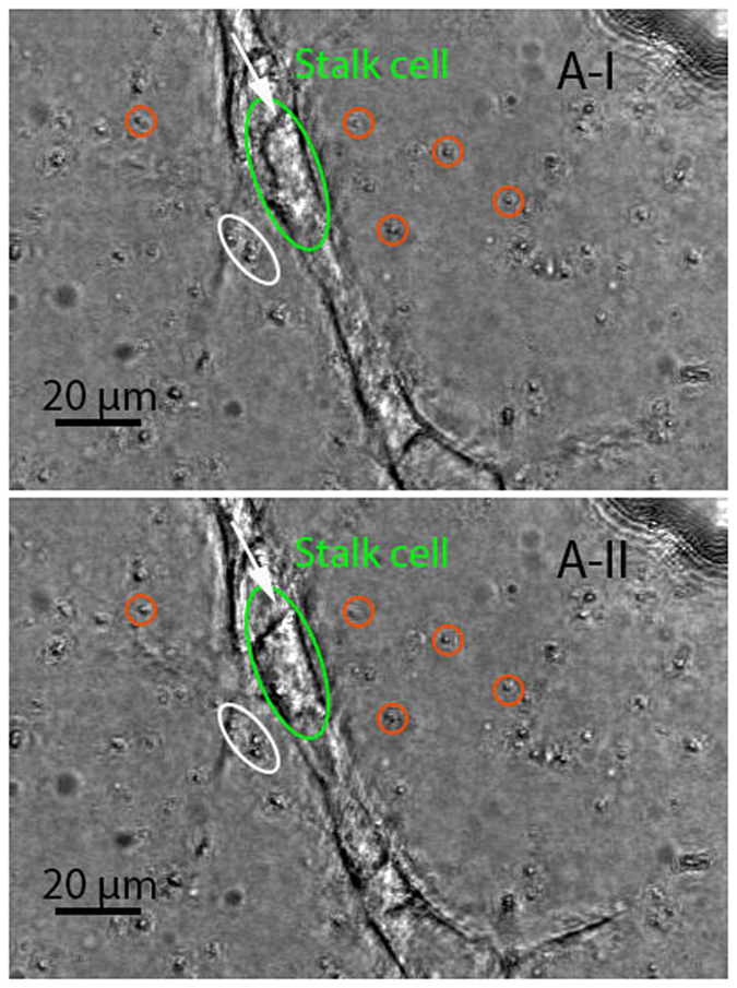
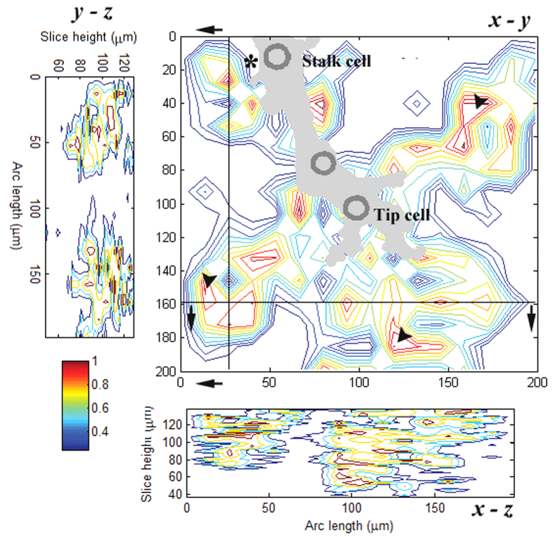
We studied the three-dimensional cell-extracellular matrix interactions of endothelial cells that form multicellular structures called sprouts. We analyzed the data collected in-situ from angiogenic sprouting experiments and identified the differentiated interaction behavior exhibited by the tip and stalk cells. Moreover, our analysis of the tip cell lamellipodia revealed the diversity in their interaction behavior under certain conditions (e.g., when the heading of a sprout is switched approximately between the long-axis direction of two different lamellipodia). This study marks the first time that new characteristics of such interactions have been identified with shape changes in the sprouts and the associated rearrangements of collagen fibers. Clear illustrations of such changes are depicted in three-dimensional views.

摘要

我们研究了形成称为芽的多细胞结构的内皮细胞的三维细胞-细胞外基质相互作用。我们分析了从血管生成芽生实验原位收集的数据,并确定了尖端细胞和茎细胞表现出的不同相互作用行为。此外,我们对尖端细胞片足的分析揭示了它们在某些条件下(例如,当一个芽的方向大约在两个不同片足的长轴方向之间切换时)相互作用行为的多样性。这项研究首次通过芽的形状变化和相关胶原纤维的重排确定了这种相互作用的新特征。这些变化的清晰图示以三维视图呈现。

https://cdn.ncbi.nlm.nih.gov/pmc/blobs/36ac/4763258/a6db49c89b75/srep21362-f11.jpg
https://cdn.ncbi.nlm.nih.gov/pmc/blobs/36ac/4763258/f13e613f73ab/srep21362-f1.jpg
https://cdn.ncbi.nlm.nih.gov/pmc/blobs/36ac/4763258/b770cc6508c1/srep21362-f2.jpg
https://cdn.ncbi.nlm.nih.gov/pmc/blobs/36ac/4763258/ec03c1630e5f/srep21362-f3.jpg
https://cdn.ncbi.nlm.nih.gov/pmc/blobs/36ac/4763258/50b51f51f084/srep21362-f4.jpg
https://cdn.ncbi.nlm.nih.gov/pmc/blobs/36ac/4763258/4548f061ab4f/srep21362-f5.jpg
https://cdn.ncbi.nlm.nih.gov/pmc/blobs/36ac/4763258/d19cf160af6e/srep21362-f6.jpg
https://cdn.ncbi.nlm.nih.gov/pmc/blobs/36ac/4763258/37287454d524/srep21362-f7.jpg
https://cdn.ncbi.nlm.nih.gov/pmc/blobs/36ac/4763258/f0157055678a/srep21362-f8.jpg
https://cdn.ncbi.nlm.nih.gov/pmc/blobs/36ac/4763258/46eaa403bd4b/srep21362-f9.jpg
https://cdn.ncbi.nlm.nih.gov/pmc/blobs/36ac/4763258/3cac9429a511/srep21362-f10.jpg
https://cdn.ncbi.nlm.nih.gov/pmc/blobs/36ac/4763258/a6db49c89b75/srep21362-f11.jpg

相似文献

1
Three-Dimensional Characterization of Mechanical Interactions between Endothelial Cells and Extracellular Matrix during Angiogenic Sprouting.血管生成芽过程中内皮细胞与细胞外基质之间机械相互作用的三维表征
Sci Rep. 2016 Feb 23;6:21362. doi: 10.1038/srep21362.
2
Matrix deformations around angiogenic sprouts correlate to sprout dynamics and suggest pulling activity.血管生成芽周围的基质变形与芽的动力学相关,并提示存在牵拉活动。
Angiogenesis. 2020 Aug;23(3):315-324. doi: 10.1007/s10456-020-09708-y. Epub 2020 Jan 29.
3
Matrix density alters zyxin phosphorylation, which limits peripheral process formation and extension in endothelial cells invading 3D collagen matrices.基质密度改变黏着斑蛋白的磷酸化,从而限制了内皮细胞在 3D 胶原基质中浸润时的周边突起的形成和延伸。
Matrix Biol. 2014 Sep;38:36-47. doi: 10.1016/j.matbio.2014.06.006. Epub 2014 Jul 16.
4
Biomimetic model to reconstitute angiogenic sprouting morphogenesis in vitro.体外重建血管生成发芽形态发生的仿生模型。
Proc Natl Acad Sci U S A. 2013 Apr 23;110(17):6712-7. doi: 10.1073/pnas.1221526110. Epub 2013 Apr 8.
5
Nonlinear optical microscopy reveals invading endothelial cells anisotropically alter three-dimensional collagen matrices.非线性光学显微镜显示,侵入的内皮细胞各向异性地改变三维胶原基质。
Exp Cell Res. 2009 Feb 1;315(3):396-410. doi: 10.1016/j.yexcr.2008.10.040. Epub 2008 Nov 6.
6
Angiogenesis-on-a-chip coupled with single-cell RNA sequencing reveals spatially differential activations of autophagy along angiogenic sprouts.血管生成芯片与单细胞 RNA 测序相结合揭示了沿着血管生成芽的自噬的空间差异激活。
Nat Commun. 2024 Jan 3;15(1):230. doi: 10.1038/s41467-023-44427-0.
7
Myosin IIA-mediated forces regulate multicellular integrity during vascular sprouting.肌球蛋白 IIA 介导的力调节血管发芽过程中的细胞间完整性。
Mol Biol Cell. 2019 Jul 22;30(16):1974-1984. doi: 10.1091/mbc.E19-02-0076. Epub 2019 Jul 18.
8
Polo-like kinase 2 regulates angiogenic sprouting and blood vessel development.Polo样激酶2调节血管生成芽和血管发育。
Dev Biol. 2015 Aug 15;404(2):49-60. doi: 10.1016/j.ydbio.2015.05.011. Epub 2015 May 22.
9
Mechanisms of vessel branching: filopodia on endothelial tip cells lead the way.血管分支的机制:内皮尖端细胞上的丝状伪足起引导作用。
Arterioscler Thromb Vasc Biol. 2009 May;29(5):639-49. doi: 10.1161/ATVBAHA.109.185165. Epub 2009 Mar 5.
10
2D data-driven stalk cell prediction model based on tip-stalk cell interaction in angiogenesis.基于血管生成中顶端-柄细胞相互作用的二维数据驱动的柄细胞预测模型。
Annu Int Conf IEEE Eng Med Biol Soc. 2013;2013:4537-40. doi: 10.1109/EMBC.2013.6610556.

引用本文的文献

1
External mechanical loading overrules cell-cell mechanical communication in sprouting angiogenesis during early bone regeneration.外部机械加载在早期骨再生过程中的发芽血管生成中胜过细胞间机械通讯。
PLoS Comput Biol. 2023 Nov 13;19(11):e1011647. doi: 10.1371/journal.pcbi.1011647. eCollection 2023 Nov.
2
Mechanical regulation of the early stages of angiogenesis.机械调控血管生成的早期阶段。
J R Soc Interface. 2022 Dec;19(197):20220360. doi: 10.1098/rsif.2022.0360. Epub 2022 Dec 7.
3
Recent advancements and future requirements in vascularization of cortical organoids.

本文引用的文献

1
A Comparative Study of Collagen Matrix Density Effect on Endothelial Sprout Formation Using Experimental and Computational Approaches.使用实验和计算方法对胶原蛋白基质密度对内皮细胞芽形成的影响进行的比较研究。
Ann Biomed Eng. 2016 Apr;44(4):929-41. doi: 10.1007/s10439-015-1416-2. Epub 2015 Aug 14.
2
Large-scale time series microscopy of neovessel growth during angiogenesis.血管生成过程中新血管生长的大规模时间序列显微镜观察。
Angiogenesis. 2015 Jul;18(3):219-32. doi: 10.1007/s10456-015-9461-x. Epub 2015 Mar 21.
3
Quantification of magnetically induced changes in ECM local apparent stiffness.
皮质类器官血管化的最新进展与未来需求
Front Bioeng Biotechnol. 2022 Nov 3;10:1048731. doi: 10.3389/fbioe.2022.1048731. eCollection 2022.
4
Bioprinted microvasculature: progressing from structure to function.生物打印微脉管:从结构到功能的进展。
Biofabrication. 2022 Feb 23;14(2). doi: 10.1088/1758-5090/ac4fb5.
5
Tissue Engineered Neurovascularization Strategies for Craniofacial Tissue Regeneration.组织工程化神经血管化策略促进颅面组织再生。
ACS Appl Bio Mater. 2022 Jan 17;5(1):20-39. doi: 10.1021/acsabm.1c00979. Epub 2021 Nov 29.
6
Mechanical Aspects of Angiogenesis.血管生成的力学方面
Cancers (Basel). 2021 Oct 5;13(19):4987. doi: 10.3390/cancers13194987.
7
3D Printing of Microgel Scaffolds with Tunable Void Fraction to Promote Cell Infiltration.3D 打印具有可调空隙率的微凝胶支架以促进细胞渗透。
Adv Healthc Mater. 2021 Sep;10(18):e2100644. doi: 10.1002/adhm.202100644. Epub 2021 Aug 3.
8
Dynamic Endothelial Stalk Cell-Matrix Interactions Regulate Angiogenic Sprout Diameter.动态内皮细胞干/祖细胞-基质相互作用调节血管生成芽的直径。
Front Bioeng Biotechnol. 2021 Mar 19;9:620128. doi: 10.3389/fbioe.2021.620128. eCollection 2021.
9
Materials for blood brain barrier modeling .用于血脑屏障建模的材料。
Mater Sci Eng R Rep. 2020 Apr;140. doi: 10.1016/j.mser.2019.100522. Epub 2020 Jan 6.
10
A multiscale model of complex endothelial cell dynamics in early angiogenesis.早期血管生成中复杂内皮细胞动力学的多尺度模型。
PLoS Comput Biol. 2021 Jan 7;17(1):e1008055. doi: 10.1371/journal.pcbi.1008055. eCollection 2021 Jan.
量化 ECM 局部表观硬度的磁诱导变化。
Biophys J. 2014 Jan 7;106(1):332-41. doi: 10.1016/j.bpj.2013.11.4459.
4
Characterization of uniaxial stiffness of extracellular matrix embedded with magnetic beads via bio-conjugation and under the influence of an external magnetic field.通过生物共轭和外加磁场的影响,对嵌入磁珠的细胞外基质的单轴弹性进行表征。
J Mech Behav Biomed Mater. 2014 Feb;30:253-65. doi: 10.1016/j.jmbbm.2013.11.012. Epub 2013 Nov 28.
5
The role of filopodia in the recognition of nanotopographies.丝状伪足在纳米形貌识别中的作用。
Sci Rep. 2013;3:1658. doi: 10.1038/srep01658.
6
Digital Volume Correlation for Study of the Mechanics of Whole Bones.用于研究全骨力学的数字体积相关技术
Procedia IUTAM. 2012;4:116-125. doi: 10.1016/j.piutam.2012.05.013.
7
Fast and robust optical flow for time-lapse microscopy using super-voxels.基于超像素的延时显微镜快速鲁棒光流算法。
Bioinformatics. 2013 Feb 1;29(3):373-80. doi: 10.1093/bioinformatics/bts706. Epub 2012 Dec 14.
8
Dynamic mechanisms of cell rigidity sensing: insights from a computational model of actomyosin networks.细胞刚性感知的动力学机制:来自肌动球蛋白网络计算模型的见解。
PLoS One. 2012;7(11):e49174. doi: 10.1371/journal.pone.0049174. Epub 2012 Nov 5.
9
VEGF and Notch in tip and stalk cell selection.血管内皮生长因子(VEGF)和 Notch 在顶端和茎细胞选择中的作用。
Cold Spring Harb Perspect Med. 2013 Jan 1;3(1):a006569. doi: 10.1101/cshperspect.a006569.
10
Group refractive index measurement of dry and hydrated type I collagen films using optical low-coherence reflectometry.使用光学低相干反射测量法对干燥和水合的I型胶原膜进行群折射率测量。
J Biomed Opt. 1996 Apr;1(2):212-6. doi: 10.1117/12.227699.