van der Kemp Wybe J M, Stehouwer Bertine L, Runge Jurgen H, Wijnen Jannie P, Nederveen Aart J, Luijten Peter R, Klomp Dennis W J
Radiology, University Medical Center Utrecht , Utrecht , Netherlands.
Radiology, Academic Medical Center , Amsterdam , Netherlands.
Front Oncol. 2016 Feb 15;6:29. doi: 10.3389/fonc.2016.00029. eCollection 2016.
The identification of the phosphodiester (PDE) (31)P MR signals in the healthy human breast at ultra-high field.
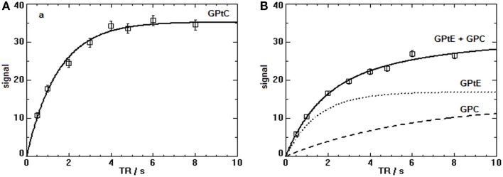
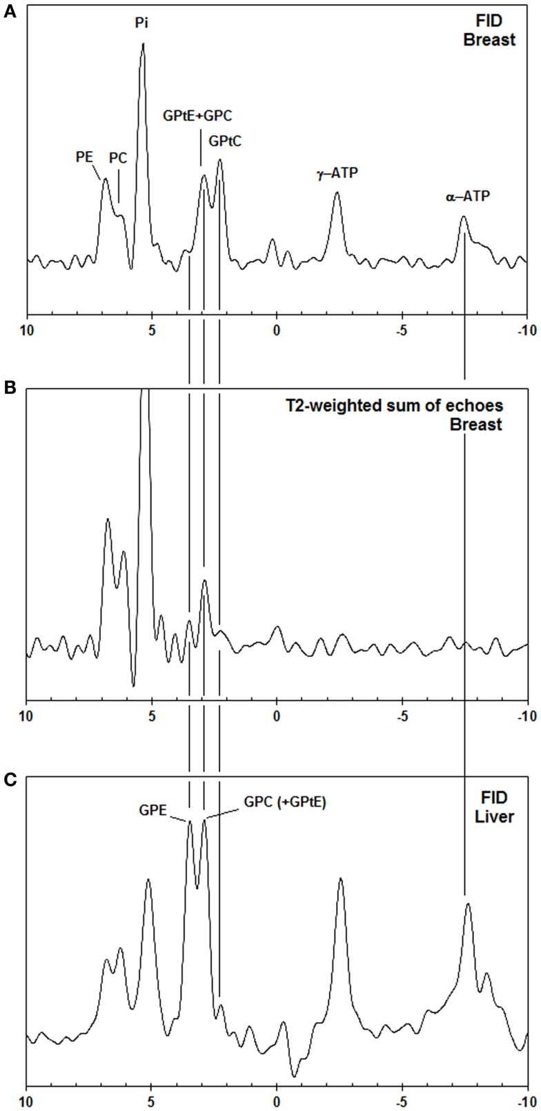
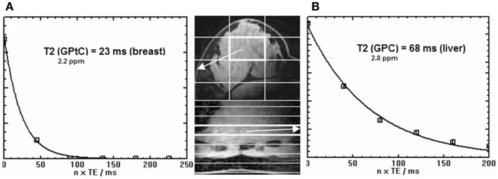
In vivo (31)P MRS measurements at 7 T of the PDE signals in the breast were performed investigating the chemical shifts, the transverse- and the longitudinal relaxation times. Chemical shifts and transverse relaxation times were compared with non-ambiguous PDE signals from the liver.
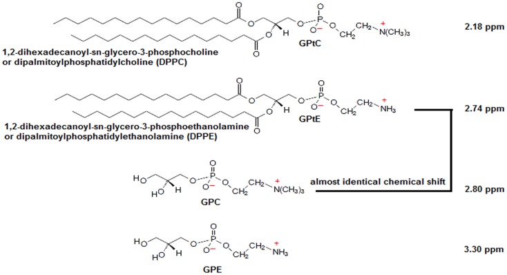
The chemical shifts of the PDE signals are shifted -0.5 ppm with respect to glycerophosphocholine (GPC) and glycerophosphoethanolamine (GPE), and the transverse and longitudinal relaxation times for these signals are a factor 3 to 4 shorter than expected for aqueous GPC and GPE.
The available experimental evidence suggests that GPC and GPE are not the main source of the PDE signals measured in fibroglandular breast tissue at 7 T. These signals may predominantly originate from mobile phospholipids.
在超高场强下识别健康人乳腺中磷酸二酯(PDE)的(31)P磁共振信号。
在7T场强下对乳腺中的PDE信号进行体内(31)P磁共振波谱测量,研究其化学位移、横向弛豫时间和纵向弛豫时间。将化学位移和横向弛豫时间与来自肝脏的明确的PDE信号进行比较。
PDE信号的化学位移相对于甘油磷酸胆碱(GPC)和甘油磷酸乙醇胺(GPE)偏移了-0.5 ppm,这些信号的横向和纵向弛豫时间比水相GPC和GPE预期的短3至4倍。
现有实验证据表明,GPC和GPE不是在7T场强下测量的纤维腺性乳腺组织中PDE信号的主要来源。这些信号可能主要源自可移动的磷脂。