• 文献检索
  • 文档翻译
  • 深度研究
  • 学术资讯
  • Suppr Zotero 插件Zotero 插件
  • 邀请有礼
  • 套餐&价格
  • 历史记录
应用&插件
Suppr Zotero 插件Zotero 插件浏览器插件Mac 客户端Windows 客户端微信小程序
定价
高级版会员购买积分包购买API积分包
服务
文献检索文档翻译深度研究API 文档MCP 服务
关于我们
关于 Suppr公司介绍联系我们用户协议隐私条款
关注我们

Suppr 超能文献

核心技术专利:CN118964589B侵权必究
粤ICP备2023148730 号-1Suppr @ 2026

文献检索

告别复杂PubMed语法,用中文像聊天一样搜索,搜遍4000万医学文献。AI智能推荐,让科研检索更轻松。

立即免费搜索

文件翻译

保留排版,准确专业,支持PDF/Word/PPT等文件格式,支持 12+语言互译。

免费翻译文档

深度研究

AI帮你快速写综述,25分钟生成高质量综述,智能提取关键信息,辅助科研写作。

立即免费体验

一种人类冠状病毒刺突蛋白的融合前结构。

Pre-fusion structure of a human coronavirus spike protein.

作者信息

Kirchdoerfer Robert N, Cottrell Christopher A, Wang Nianshuang, Pallesen Jesper, Yassine Hadi M, Turner Hannah L, Corbett Kizzmekia S, Graham Barney S, McLellan Jason S, Ward Andrew B

机构信息

Department of Integrative Structural and Computational Biology, The Scripps Research Institute, 10550 North Torrey Pines Road, La Jolla, California 92037, USA.

Department of Biochemistry, Geisel School of Medicine at Dartmouth, Hanover, New Hampshire 03755, USA.

出版信息

Nature. 2016 Mar 3;531(7592):118-21. doi: 10.1038/nature17200.

DOI:10.1038/nature17200
PMID:26935699
原文链接:https://pmc.ncbi.nlm.nih.gov/articles/PMC4860016/
Abstract

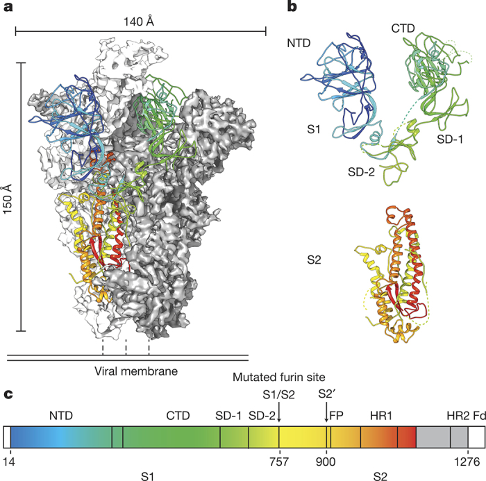
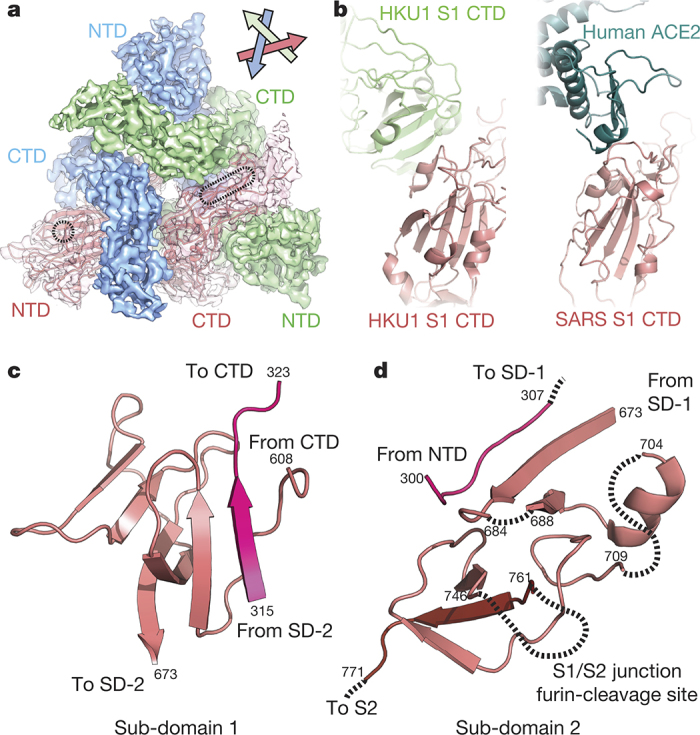
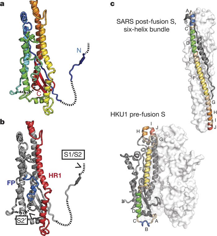
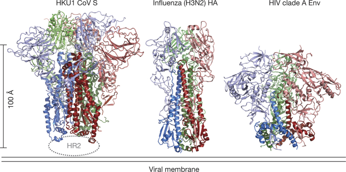
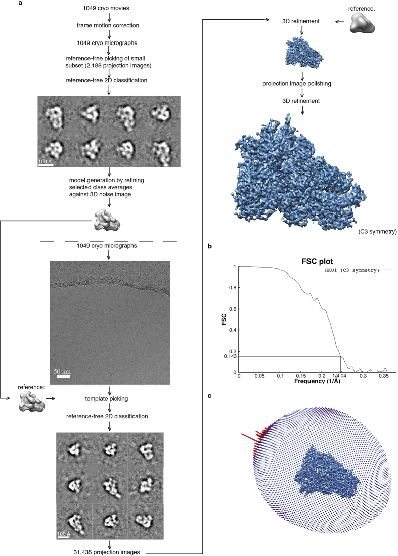
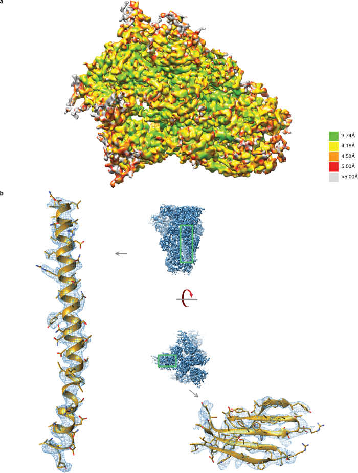
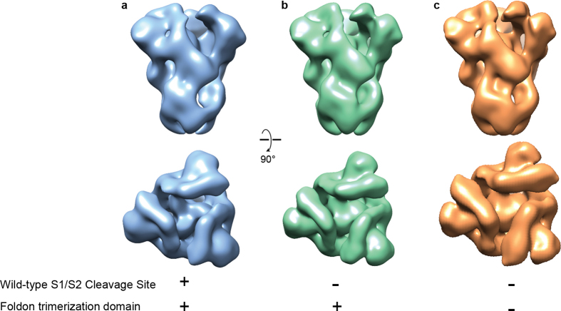
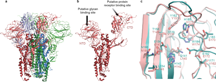
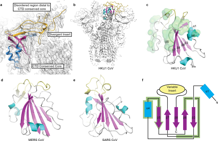
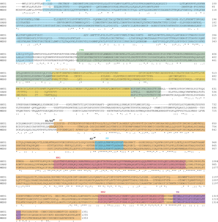
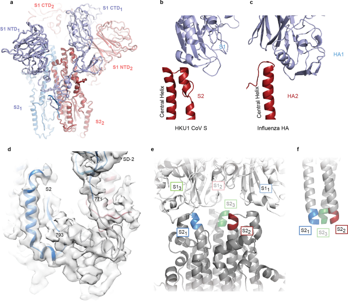
HKU1 is a human betacoronavirus that causes mild yet prevalent respiratory disease, and is related to the zoonotic SARS and MERS betacoronaviruses, which have high fatality rates and pandemic potential. Cell tropism and host range is determined in part by the coronavirus spike (S) protein, which binds cellular receptors and mediates membrane fusion. As the largest known class I fusion protein, its size and extensive glycosylation have hindered structural studies of the full ectodomain, thus preventing a molecular understanding of its function and limiting development of effective interventions. Here we present the 4.0 Å resolution structure of the trimeric HKU1 S protein determined using single-particle cryo-electron microscopy. In the pre-fusion conformation, the receptor-binding subunits, S1, rest above the fusion-mediating subunits, S2, preventing their conformational rearrangement. Surprisingly, the S1 C-terminal domains are interdigitated and form extensive quaternary interactions that occlude surfaces known in other coronaviruses to bind protein receptors. These features, along with the location of the two protease sites known to be important for coronavirus entry, provide a structural basis to support a model of membrane fusion mediated by progressive S protein destabilization through receptor binding and proteolytic cleavage. These studies should also serve as a foundation for the structure-based design of betacoronavirus vaccine immunogens.

摘要

HKU1是一种人类β冠状病毒,可引起轻微但普遍的呼吸道疾病,并且与具有高致死率和大流行潜力的人畜共患的严重急性呼吸综合征(SARS)和中东呼吸综合征(MERS)β冠状病毒相关。细胞嗜性和宿主范围部分由冠状病毒刺突(S)蛋白决定,该蛋白结合细胞受体并介导膜融合。作为已知最大的I类融合蛋白,其大小和广泛的糖基化阻碍了对完整胞外域的结构研究,从而妨碍了对其功能的分子理解,并限制了有效干预措施的开发。在此,我们展示了使用单颗粒冷冻电子显微镜测定的三聚体HKU1 S蛋白的4.0埃分辨率结构。在融合前构象中,受体结合亚基S1位于融合介导亚基S2上方,阻止其构象重排。令人惊讶的是,S1 C末端结构域相互交错并形成广泛的四级相互作用,从而封闭了其他冠状病毒中已知的与蛋白质受体结合的表面。这些特征,连同已知对冠状病毒进入很重要的两个蛋白酶位点的位置,为支持一种通过受体结合和蛋白水解切割使S蛋白逐步不稳定从而介导膜融合的模型提供了结构基础。这些研究也应为基于结构的β冠状病毒疫苗免疫原设计奠定基础。

https://cdn.ncbi.nlm.nih.gov/pmc/blobs/7959/7095257/eb199d940d92/41586_2016_Article_BFnature17200_Fig13_ESM.jpg
https://cdn.ncbi.nlm.nih.gov/pmc/blobs/7959/7095257/3a7cb605c3d4/41586_2016_Article_BFnature17200_Fig1_HTML.jpg
https://cdn.ncbi.nlm.nih.gov/pmc/blobs/7959/7095257/e96050f49cc8/41586_2016_Article_BFnature17200_Fig2_HTML.jpg
https://cdn.ncbi.nlm.nih.gov/pmc/blobs/7959/7095257/b68075c57689/41586_2016_Article_BFnature17200_Fig3_HTML.jpg
https://cdn.ncbi.nlm.nih.gov/pmc/blobs/7959/7095257/3a801ab017f7/41586_2016_Article_BFnature17200_Fig4_HTML.jpg
https://cdn.ncbi.nlm.nih.gov/pmc/blobs/7959/7095257/cbe5db1c95ad/41586_2016_Article_BFnature17200_Fig5_ESM.jpg
https://cdn.ncbi.nlm.nih.gov/pmc/blobs/7959/7095257/8dc08e9d914b/41586_2016_Article_BFnature17200_Fig6_ESM.jpg
https://cdn.ncbi.nlm.nih.gov/pmc/blobs/7959/7095257/2c800cedbd87/41586_2016_Article_BFnature17200_Fig7_ESM.jpg
https://cdn.ncbi.nlm.nih.gov/pmc/blobs/7959/7095257/b55c08a760dc/41586_2016_Article_BFnature17200_Fig8_ESM.jpg
https://cdn.ncbi.nlm.nih.gov/pmc/blobs/7959/7095257/f375363397de/41586_2016_Article_BFnature17200_Fig9_ESM.jpg
https://cdn.ncbi.nlm.nih.gov/pmc/blobs/7959/7095257/29d712e96625/41586_2016_Article_BFnature17200_Fig10_ESM.jpg
https://cdn.ncbi.nlm.nih.gov/pmc/blobs/7959/7095257/3d75ca6aa072/41586_2016_Article_BFnature17200_Fig11_ESM.jpg
https://cdn.ncbi.nlm.nih.gov/pmc/blobs/7959/7095257/37dc0015fded/41586_2016_Article_BFnature17200_Fig12_ESM.jpg
https://cdn.ncbi.nlm.nih.gov/pmc/blobs/7959/7095257/eb199d940d92/41586_2016_Article_BFnature17200_Fig13_ESM.jpg

相似文献

1
Pre-fusion structure of a human coronavirus spike protein.一种人类冠状病毒刺突蛋白的融合前结构。
Nature. 2016 Mar 3;531(7592):118-21. doi: 10.1038/nature17200.
2
Structure of mouse coronavirus spike protein complexed with receptor reveals mechanism for viral entry.鼠冠状病毒刺突蛋白复合物与受体的结构揭示了病毒进入的机制。
PLoS Pathog. 2020 Mar 9;16(3):e1008392. doi: 10.1371/journal.ppat.1008392. eCollection 2020 Mar.
3
Cryo-EM structure of the SARS coronavirus spike glycoprotein in complex with its host cell receptor ACE2.SARS 冠状病毒刺突糖蛋白与其宿主细胞受体 ACE2 复合物的冷冻电镜结构。
PLoS Pathog. 2018 Aug 13;14(8):e1007236. doi: 10.1371/journal.ppat.1007236. eCollection 2018 Aug.
4
Receptor binding and priming of the spike protein of SARS-CoV-2 for membrane fusion.SARS-CoV-2 刺突蛋白的受体结合和引发膜融合。
Nature. 2020 Dec;588(7837):327-330. doi: 10.1038/s41586-020-2772-0. Epub 2020 Sep 17.
5
Biochemical Analysis of Coronavirus Spike Glycoprotein Conformational Intermediates during Membrane Fusion.冠状病毒刺突糖蛋白在膜融合过程中构象中间体的生化分析。
J Virol. 2019 Sep 12;93(19). doi: 10.1128/JVI.00785-19. Print 2019 Oct 1.
6
Cryo-Electron Microscopy Structure of Porcine Deltacoronavirus Spike Protein in the Prefusion State.冷冻电镜下猪德尔塔冠状病毒刺突蛋白三聚体的预融合构象
J Virol. 2018 Feb 15;92(4):e01556-17. doi: 10.1128/JVI.01556-17.
7
Coronavirus Spike Protein and Tropism Changes.冠状病毒刺突蛋白与嗜性变化
Adv Virus Res. 2016;96:29-57. doi: 10.1016/bs.aivir.2016.08.004. Epub 2016 Sep 13.
8
Cryo-EM analysis of the post-fusion structure of the SARS-CoV spike glycoprotein.冷冻电镜分析 SARS-CoV 刺突糖蛋白的融合后结构。
Nat Commun. 2020 Jul 17;11(1):3618. doi: 10.1038/s41467-020-17371-6.
9
The 3.1-Angstrom Cryo-electron Microscopy Structure of the Porcine Epidemic Diarrhea Virus Spike Protein in the Prefusion Conformation.猪流行性腹泻病毒刺突蛋白在融合前构象的 3.1 埃冷冻电镜结构。
J Virol. 2019 Nov 13;93(23). doi: 10.1128/JVI.00923-19. Print 2019 Dec 1.
10
Cryo-electron microscopy structure of a coronavirus spike glycoprotein trimer.冠状病毒刺突糖蛋白三聚体的冷冻电子显微镜结构
Nature. 2016 Mar 3;531(7592):114-117. doi: 10.1038/nature16988. Epub 2016 Feb 8.

引用本文的文献

1
Structural insights into TLR4 activation by SARS-CoV-2 spike protein: implications for inflammatory response modulation.SARS-CoV-2刺突蛋白激活TLR4的结构见解:对炎症反应调节的影响
Mol Divers. 2025 Sep 13. doi: 10.1007/s11030-025-11347-8.
2
Cross-neutralization ability of anti-MERS-CoV monoclonal antibodies against a variety of merbecoviruses.抗中东呼吸综合征冠状病毒单克隆抗体对多种默贝病毒的交叉中和能力。
Front Microbiol. 2025 Jul 16;16:1593095. doi: 10.3389/fmicb.2025.1593095. eCollection 2025.
3
Merbecovirus S2 subunit vaccines elicit cross reactive antibodies and provide partial protection against MERS coronavirus.

本文引用的文献

1
Structure and stabilization of the Hendra virus F glycoprotein in its prefusion form.亨德拉病毒融合前形式的F糖蛋白的结构与稳定性
Proc Natl Acad Sci U S A. 2016 Jan 26;113(4):1056-61. doi: 10.1073/pnas.1523303113. Epub 2015 Dec 28.
2
Crystal Structure of the Pre-fusion Nipah Virus Fusion Glycoprotein Reveals a Novel Hexamer-of-Trimers Assembly.融合前尼帕病毒融合糖蛋白的晶体结构揭示了一种新型三聚体六聚体组装形式。
PLoS Pathog. 2015 Dec 8;11(12):e1005322. doi: 10.1371/journal.ppat.1005322. eCollection 2015 Dec.
3
Complete epitopes for vaccine design derived from a crystal structure of the broadly neutralizing antibodies PGT128 and 8ANC195 in complex with an HIV-1 Env trimer.
默贝科病毒S2亚基疫苗可引发交叉反应性抗体,并对中东呼吸综合征冠状病毒提供部分保护。
Npj Viruses. 2025 Jul 29;3(1):60. doi: 10.1038/s44298-025-00142-9.
4
Virus glycoprotein nanodisc platform for vaccine design.用于疫苗设计的病毒糖蛋白纳米盘平台
bioRxiv. 2025 Jun 16:2025.05.02.651272. doi: 10.1101/2025.05.02.651272.
5
Tailoring the adjuvanticity of lipid nanoparticles by PEG lipid ratio and phospholipid modifications.通过聚乙二醇脂质比例和磷脂修饰来调整脂质纳米颗粒的佐剂活性。
Nat Nanotechnol. 2025 Jun 23. doi: 10.1038/s41565-025-01958-5.
6
Complex Donuts: Small Variations in DNA Sequence Dictate Pathway Complexity in DNA Nanotoroids.复杂的甜甜圈:DNA序列中的微小变异决定了DNA纳米环的途径复杂性。
Angew Chem Int Ed Engl. 2025 Aug 11;64(33):e202501441. doi: 10.1002/anie.202501441. Epub 2025 Jul 4.
7
Synthesis and anti-SARS-CoV-2 potential of novel coumarin hybrids: a combined wet/dry lab approach targeting M, Nsp15 and spike protein.新型香豆素杂化物的合成及其抗SARS-CoV-2潜力:一种针对M、Nsp15和刺突蛋白的湿/干实验室联合方法
RSC Adv. 2025 Jun 3;15(23):18577-18592. doi: 10.1039/d5ra02615f. eCollection 2025 May 29.
8
A neutralizing human antibody induces movement of the HCoV-229E receptor binding domain.一种中和性人抗体诱导人冠状病毒229E受体结合域的移动。
bioRxiv. 2025 May 17:2025.05.14.654097. doi: 10.1101/2025.05.14.654097.
9
Centrifugation-Based Purification Protocol Optimization Enhances Structural Preservation of Nucleopolyhedrovirus Budded Virion Envelopes.基于离心的纯化方案优化可增强核多角体病毒出芽病毒粒子包膜的结构保存。
Insects. 2025 Apr 17;16(4):424. doi: 10.3390/insects16040424.
10
Elicitation of liver-stage immunity by nanoparticle immunogens displaying P. falciparum CSP-derived antigens.展示恶性疟原虫环子孢子蛋白(CSP)衍生抗原的纳米颗粒免疫原引发肝期免疫。
NPJ Vaccines. 2025 May 5;10(1):87. doi: 10.1038/s41541-025-01140-x.
源自广泛中和抗体PGT128和8ANC195与HIV-1包膜三聚体复合物晶体结构的用于疫苗设计的完整表位。
Acta Crystallogr D Biol Crystallogr. 2015 Oct;71(Pt 10):2099-108. doi: 10.1107/S1399004715013917. Epub 2015 Sep 26.
4
EMRinger: side chain-directed model and map validation for 3D cryo-electron microscopy.EMRinger:用于三维冷冻电子显微镜的侧链导向模型与图谱验证
Nat Methods. 2015 Oct;12(10):943-6. doi: 10.1038/nmeth.3541. Epub 2015 Aug 17.
5
Identification of the Receptor-Binding Domain of the Spike Glycoprotein of Human Betacoronavirus HKU1.人乙型冠状病毒HKU1刺突糖蛋白受体结合域的鉴定
J Virol. 2015 Sep;89(17):8816-27. doi: 10.1128/JVI.03737-14. Epub 2015 Jun 17.
6
Human Coronavirus HKU1 Spike Protein Uses O-Acetylated Sialic Acid as an Attachment Receptor Determinant and Employs Hemagglutinin-Esterase Protein as a Receptor-Destroying Enzyme.人冠状病毒HKU1刺突蛋白以O-乙酰化唾液酸作为附着受体决定簇,并利用血凝素酯酶蛋白作为受体破坏酶。
J Virol. 2015 Jul;89(14):7202-13. doi: 10.1128/JVI.00854-15. Epub 2015 Apr 29.
7
Atomic-accuracy models from 4.5-Å cryo-electron microscopy data with density-guided iterative local refinement.基于4.5埃冷冻电子显微镜数据并采用密度引导迭代局部优化的原子精度模型。
Nat Methods. 2015 Apr;12(4):361-365. doi: 10.1038/nmeth.3286. Epub 2015 Feb 23.
8
Clustal omega.Clustal欧米伽
Curr Protoc Bioinformatics. 2014 Dec 12;48:3.13.1-3.13.16. doi: 10.1002/0471250953.bi0313s48.
9
Host cell proteases: Critical determinants of coronavirus tropism and pathogenesis.宿主细胞蛋白酶:冠状病毒嗜性和发病机制的关键决定因素。
Virus Res. 2015 Apr 16;202:120-34. doi: 10.1016/j.virusres.2014.11.021. Epub 2014 Nov 22.
10
Receptor recognition mechanisms of coronaviruses: a decade of structural studies.冠状病毒的受体识别机制:十年结构研究
J Virol. 2015 Feb;89(4):1954-64. doi: 10.1128/JVI.02615-14. Epub 2014 Nov 26.