de Boer Peter, Bleeker Maaike C G, Spijkerboer Anje M, van de Schoot Agustinus J A J, Bipat Shandra, Buist Marrije R, Rasch Coen R N, Stoker Jaap, Stalpers Lukas J A
Department of Radiation Oncology, Academic Medical Centre (AMC), University of Amsterdam (UvA), Meibergdreef 9, 1105 AZ Amsterdam, The Netherlands.
Department of Pathology, AMC, UvA, Meibergdreef 9, 1105 AZ Amsterdam, The Netherlands.
Eur J Radiol Open. 2015 Jul 26;2:111-7. doi: 10.1016/j.ejro.2015.07.001. eCollection 2015.
To assess the reliability of magnetic resonance imaging (MRI) for evaluation of craniocaudal tumour extension by comparing the craniocaudal tumour extension on the pre-operative MRI and post-operative hysterectomy specimen in patients with early stage uterine cervical cancer.
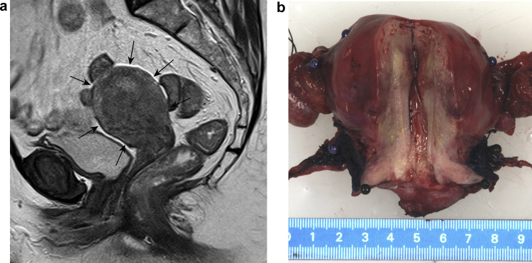
After approval of the institutional review board was acquired, pre-operative MRI and hysterectomy specimen of 21 women with early stage cervical cancer were re-evaluated. The craniocaudal extension on MRI was measured separately by two experienced radiologists and compared with corresponding measurements from the hysterectomy specimen, which were re-evaluated by an experienced pathologist.
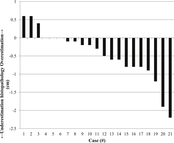
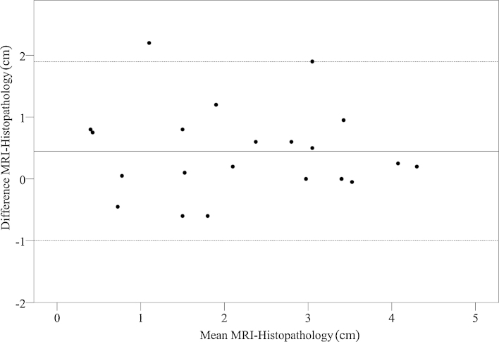
Median craniocaudal extension of uterine cervical cancer on MRI was slightly smaller compared to histopathology (2.1 cm vs. 2.5 cm). The median underestimation was 0.4 cm (range -0.6 cm to 2.2 cm, mean 0.4 cm, standard deviation (SD) ±0.7 cm); Pearson's correlation was 0.83 (p < 0.001). In two patients (9%) MRI underestimated tumour craniocaudal extension by more than 1.8 cm.
MRI represents the histopathological craniocaudal tumour extension in the majority of patients with early stage uterine cervical cancer, but with a systematic small underestimation of the real craniocaudal tumour extension.
通过比较早期子宫颈癌患者术前磁共振成像(MRI)和术后子宫切除标本的颅尾向肿瘤延伸情况,评估MRI评估颅尾向肿瘤延伸的可靠性。
获得机构审查委员会批准后,对21例早期宫颈癌患者的术前MRI和子宫切除标本进行重新评估。由两名经验丰富的放射科医生分别测量MRI上的颅尾向延伸,并与由经验丰富的病理学家重新评估的子宫切除标本的相应测量值进行比较。
与组织病理学相比,子宫颈癌在MRI上的颅尾向延伸中位数略小(2.1厘米对2.5厘米)。中位数低估为0.4厘米(范围-0.6厘米至2.2厘米,平均0.4厘米,标准差(SD)±0.7厘米);Pearson相关性为0.83(p <0.001)。在两名患者(9%)中,MRI对肿瘤颅尾向延伸的低估超过1.8厘米。
MRI在大多数早期子宫颈癌患者中可反映组织病理学上的颅尾向肿瘤延伸,但对实际颅尾向肿瘤延伸存在系统性的轻微低估。