Lee Jung-Hwan, Kang Min-Sil, Mahapatra Chinmaya, Kim Hae-Won
Institute of Tissue Regeneration Engineering (ITREN), Dankook University, Cheonan 31116, Republic of Korea.
Department of Nanobiomedical Science and BK21 PLUS NBM Global Research, Center for Regenerative Medicine, Dankook University, Cheonan 31116, Republic of Korea.
PLoS One. 2016 Mar 14;11(3):e0150727. doi: 10.1371/journal.pone.0150727. eCollection 2016.
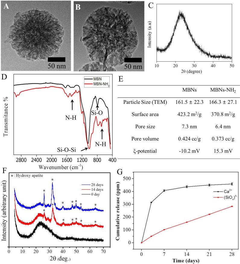
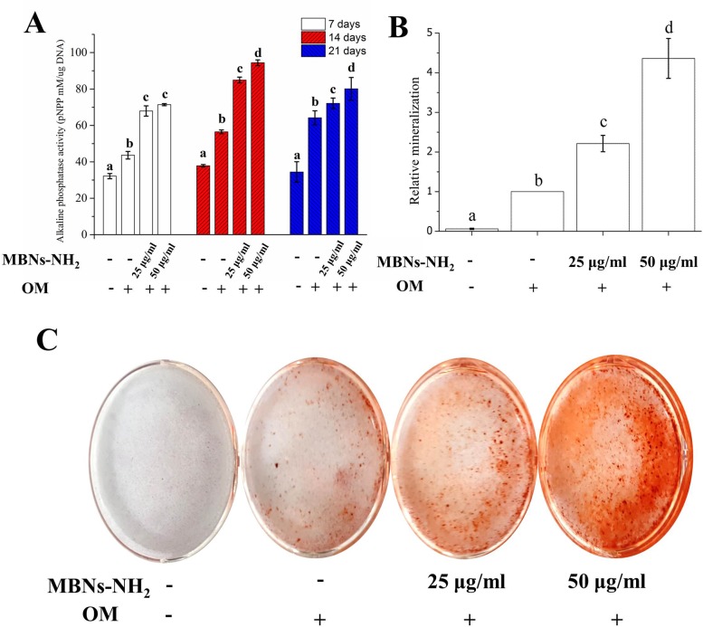
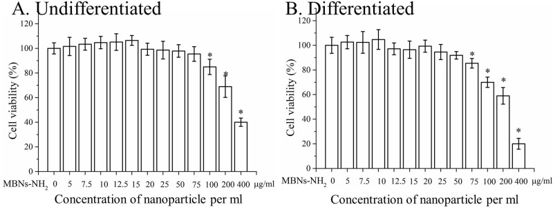
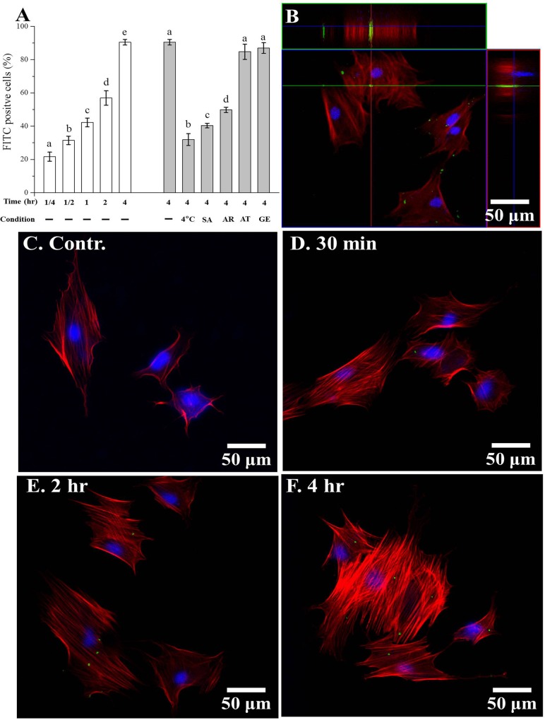
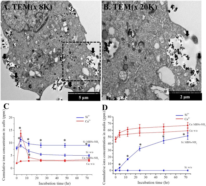
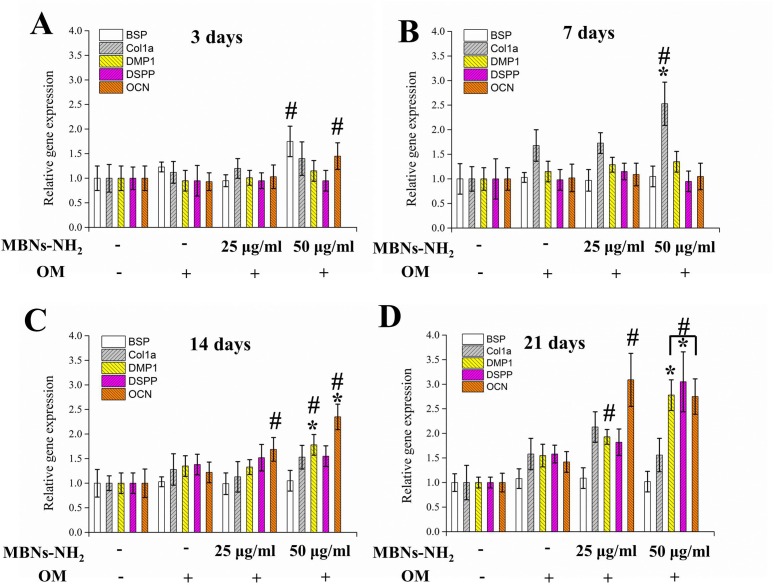
Mesoporous bioactive nanoparticles (MBNs) have been developed as promising additives to various types of bone or dentin regenerative material. However, biofunctionality of MBNs as dentin regenerative additive to dental materials have rarely been studied. We investigated the uptake efficiency of MBNs-NH2 with their endocytosis pathway and the role of MBNs-NH2 in odontogenic differentiation to clarify inherent biofunctionality. MBNs were fabricated by sol-gel synthesis, and 3% APTES was used to aminate these nanoparticles (MBNs-NH2) to reverse their charge from negative to positive. To characterize the MBNs-NH2, TEM, XRD, FTIR, zeta(ξ)-potential measurements, and Brunauer-Emmett-Teller analysis were performed. After primary cultured rat dental pulp stem cells (rDPSCs) were incubated with various concentrations of MBNs-NH2, stem cell viability (24 hours) with or without differentiated media, internalization of MBNs-NH2 in rDPSCs (4 hours) via specific endocytosis pathway, intra or extracellular ion concentration and odontoblastic differentiation (28 days) were investigated. Incubation with up to 50 μg/mL of MBNs-NH2 had no effect on rDPSCs viability with differentiated media (p>0.05). The internalization of MBNs-NH2 in rDPSCs was determined about 92% after 4 hours of incubation. Uptake was significantly decreased with ATP depletion and after 1 hour of pre-treatment with the inhibitor of macropinocytosis (p<0.05). There was significant increase of intracellular Ca and Si ion concentration in MBNs-NH2 treated cells compared to no-treated counterpart (p<0.05). The expression of odontogenic-related genes (BSP, COL1A, DMP-1, DSPP, and OCN) and the capacity for biomineralization (based on alkaline phosphatase activity and alizarin red staining) were significantly upregulated with MBNs-NH2. These results indicate that MBNs-NH2 induce odontogenic differentiation of rDPSCs and may serve as a potential dentin regenerative additive to dental material for promoting odontoblast differentiation.
介孔生物活性纳米颗粒(MBNs)已被开发为各类骨或牙本质再生材料中颇具前景的添加剂。然而,MBNs作为牙科材料中牙本质再生添加剂的生物功能却鲜有研究。我们研究了氨基化介孔生物活性纳米颗粒(MBNs-NH2)的摄取效率及其内吞途径,以及MBNs-NH2在牙源性分化中的作用,以阐明其内在生物功能。通过溶胶-凝胶合成法制备MBNs,并使用3%的3-氨丙基三乙氧基硅烷(APTES)对这些纳米颗粒进行胺化处理(MBNs-NH2),使其电荷从负变为正。为了表征MBNs-NH2,进行了透射电子显微镜(TEM)、X射线衍射(XRD)、傅里叶变换红外光谱(FTIR)、zeta(ξ)电位测量以及布鲁诺尔-埃米特-泰勒(Brunauer-Emmett-Teller)分析。将原代培养的大鼠牙髓干细胞(rDPSCs)与不同浓度的MBNs-NH2孵育后,研究了有无分化培养基时的干细胞活力(24小时)、MBNs-NH2通过特定内吞途径在rDPSCs中的内化情况(约4小时)、细胞内或细胞外离子浓度以及成牙本质细胞分化情况(约28天)。用高达50μg/mL的MBNs-NH2孵育对添加分化培养基的rDPSCs活力没有影响(p>0.05)。孵育约4小时后,rDPSCs中MBNs-NH2的内化率约为92%。ATP耗竭以及用巨胞饮作用抑制剂预处理1小时后,摄取量显著降低(p<0.05)。与未处理的细胞相比,经MBNs-NH2处理的细胞内钙和硅离子浓度显著增加(p<0.05)。MBNs-NH2显著上调了牙源性相关基因(骨涎蛋白(BSP)、I型胶原蛋白(COL1A)、牙本质基质蛋白-1(DMP-1)、牙本质涎磷蛋白(DSPP)和骨钙素(OCN))的表达以及生物矿化能力(基于碱性磷酸酶活性和茜素红染色)。这些结果表明,MBNs-NH2可诱导rDPSCs的牙源性分化,并可能作为牙科材料潜在的牙本质再生添加剂,以促进成牙本质细胞分化。