Fabregat-Andres Oscar, Paredes Federico, Monsalve María, Milara Javier, Ridocci-Soriano Francisco, Gonzalez-Hervas Sonia, Mena Armando, Facila Lorenzo, Hornero Fernando, Morell Salvador, Martinez-Leon Juan, Cortijo Julio
Cardiovascular Institute, General University Hospital, Valencia-Spain.
Research Foundation, General University Hospital, Valencia-Spain.
Anatol J Cardiol. 2016 Aug;16(8):622-629. doi: 10.5152/AnatolJCardiol.2015.6466. Epub 2015 Nov 26.
Peroxisome proliferator-activated receptor-γ coactivator-1α (PGC-1α) is a transcriptional coactivator that has been proposed to play a protective role in mouse models of cardiac ischemia and heart failure, suggesting that PGC-1α could be relevant as a prognostic marker. Our previous studies showed that the estimation of peripheral mRNA PGC-1α expression was feasible and that its induction correlated with the extent of myocardial necrosis and left ventricular remodeling in patients with myocardial infarction. In this study, we sought to determine if the myocardial and peripheral expressions of PGC-1α are well correlated and to analyze the variability of PGC-1α expression depending on the prevalence of some metabolic disorders.
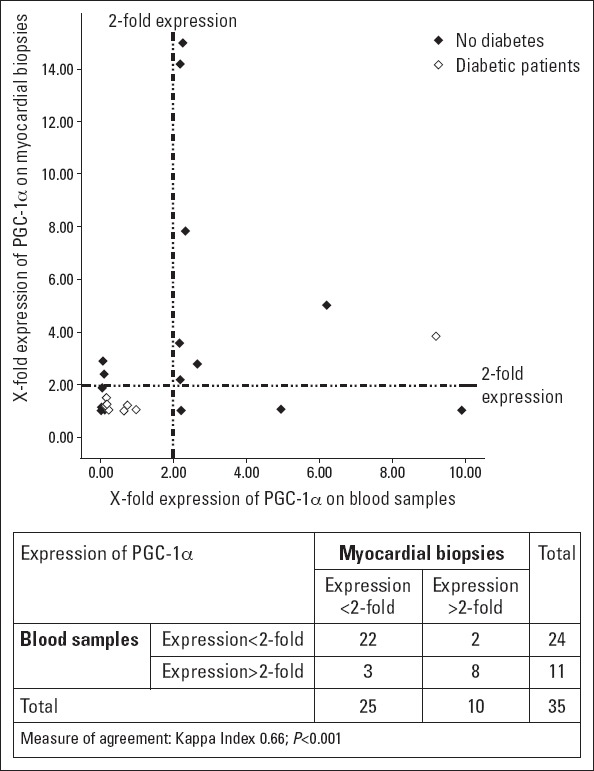
This was a cohort of 35 consecutive stable heart failure patients with severe aortic stenosis who underwent an elective aortic valve replacement surgery. mRNA PGC-1α expression was simultaneously determined from myocardial biopsy specimens and blood samples obtained during surgery by quantitative PCR, and a correlation between samples was made using the Kappa index. Patients were divided into two groups according to the detection of baseline expression levels of PGC-1α in blood samples, and comparisons between both groups were made by chi-square test or unpaired Student's t-test as appropriate.
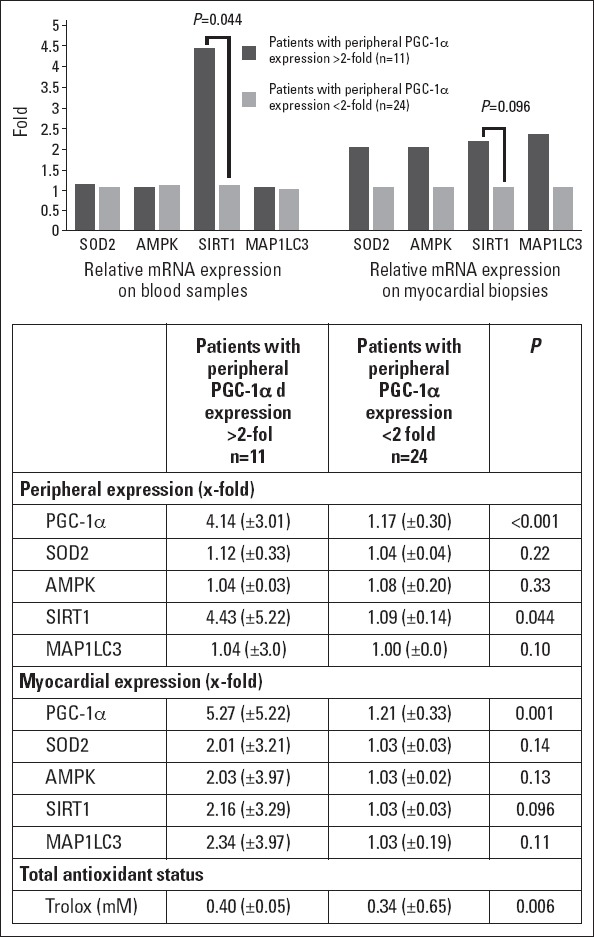
Based on myocardial biopsies, we found that mRNA PGC-1α expression in blood samples showed a statistically significant correlation with myocardial expression (Kappa index 0.66, p<0.001). The presence of higher systemic PGC-1α expression was associated with a greater expression of some target genes such as silent information regulator 2 homolog-1 (x-fold expression in blood samples: 4.43±5.22 vs. 1.09±0.14, p=0.044) and better antioxidant status in these patients (concentration of Trolox: 0.40±0.05 vs. 0.34±0.65, p=0.006).
Most patients with higher peripheral expression also had increased myocardial expression, so we conclude that the non-invasive estimation of mRNA PGC-1α expression from blood samples provides a good approach of the constitutive status of the mitochondrial protection system regulated by PGC-1α and that this could be used as prognostic indicator in cardiovascular disease.
过氧化物酶体增殖物激活受体γ共激活因子1α(PGC-1α)是一种转录共激活因子,在心脏缺血和心力衰竭的小鼠模型中被认为具有保护作用,这表明PGC-1α可能作为一种预后标志物。我们之前的研究表明,评估外周血mRNA PGC-1α表达是可行的,并且其诱导与心肌梗死患者的心肌坏死程度和左心室重构相关。在本研究中,我们试图确定PGC-1α的心肌和外周血表达是否具有良好的相关性,并分析PGC-1α表达的变异性与某些代谢紊乱患病率之间的关系。
这是一组连续35例患有严重主动脉瓣狭窄的稳定心力衰竭患者,他们接受了择期主动脉瓣置换手术。通过定量PCR同时测定手术期间获取的心肌活检标本和血样中的mRNA PGC-1α表达,并使用Kappa指数对样本进行相关性分析。根据血样中PGC-1α基线表达水平的检测结果将患者分为两组,并根据情况采用卡方检验或非配对学生t检验对两组进行比较。
基于心肌活检,我们发现血样中的mRNA PGC-1α表达与心肌表达具有统计学意义的相关性(Kappa指数0.66,p<0.001)。全身PGC-1α表达较高与一些靶基因的更高表达相关,如沉默信息调节因子2同源物1(血样中的x倍表达:4.43±5.22 vs. 1.09±0.14,p=0.044),并且这些患者的抗氧化状态更好(Trolox浓度:0.40±0.05 vs. 0.34±0.65,p=0.006)。
大多数外周血表达较高的患者心肌表达也增加,因此我们得出结论,从血样中对mRNA PGC-1α表达进行非侵入性评估为PGC-1α调节的线粒体保护系统的组成状态提供了一种良好的方法,并且这可作为心血管疾病的预后指标。