Institute for Community Medicine, University of Greifswald, Greifswald, Germany.
Department of Radiology, Center for Biomedical Image Computing and Analytics, University of Pennsylvania, Philadelphia, PA, USA.
Transl Psychiatry. 2016 Apr 5;6(4):e775. doi: 10.1038/tp.2016.39.
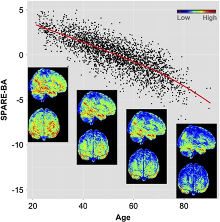
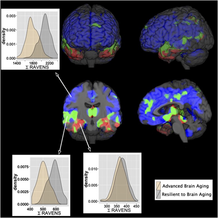
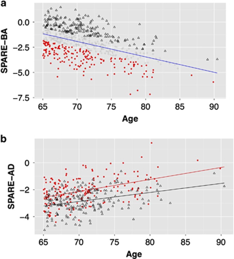
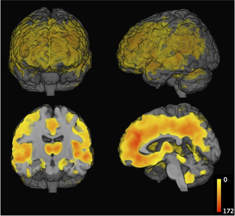
We systematically compared structural imaging patterns of advanced brain aging (ABA) in the general-population, herein defined as significant deviation from typical BA to those found in Alzheimer disease (AD). The hypothesis that ABA would show different patterns of structural change compared with those found in AD was tested via advanced pattern analysis methods. In particular, magnetic resonance images of 2705 participants from the Study of Health in Pomerania (aged 20-90 years) were analyzed using an index that captures aging atrophy patterns (Spatial Pattern of Atrophy for Recognition of BA (SPARE-BA)), and an index previously shown to capture atrophy patterns found in clinical AD (Spatial Patterns of Abnormality for Recognition of Early Alzheimer's Disease (SPARE-AD)). We studied the association between these indices and risk factors, including an AD polygenic risk score. Finally, we compared the ABA-associated atrophy with typical AD-like patterns. We observed that SPARE-BA had significant association with: smoking (P<0.05), anti-hypertensive (P<0.05), anti-diabetic drug use (men P<0.05, women P=0.06) and waist circumference for the male cohort (P<0.05), after adjusting for age. Subjects with ABA had spatially extensive gray matter loss in the frontal, parietal and temporal lobes (false-discovery-rate-corrected q<0.001). ABA patterns of atrophy were partially overlapping with, but notably deviating from those typically found in AD. Subjects with ABA had higher SPARE-AD values; largely due to the partial spatial overlap of associated patterns in temporal regions. The AD polygenic risk score was significantly associated with SPARE-AD but not with SPARE-BA. Our findings suggest that ABA is likely characterized by pathophysiologic mechanisms that are distinct from, or only partially overlapping with those of AD.
我们系统地比较了一般人群中高级脑衰老(ABA)的结构成像模式,将其定义为与典型脑衰老相比存在显著偏差的模式,与阿尔茨海默病(AD)中发现的模式进行比较。通过先进的模式分析方法,测试了 ABA 会表现出与 AD 中发现的模式不同的结构变化模式的假设。特别是,使用捕获老化萎缩模式的指数(用于识别 BA 的空间萎缩模式指数(SPARE-BA))分析了来自波美拉尼亚健康研究(年龄 20-90 岁)的 2705 名参与者的磁共振图像,以及以前显示捕获在临床 AD 中发现的萎缩模式的指数(用于识别早期阿尔茨海默病的异常空间模式(SPARE-AD))。我们研究了这些指数与风险因素之间的关联,包括 AD 多基因风险评分。最后,我们比较了与 ABA 相关的萎缩与典型的 AD 样模式。我们观察到,SPARE-BA 与以下因素显著相关:吸烟(P<0.05)、抗高血压药物(P<0.05)、抗糖尿病药物(男性 P<0.05,女性 P=0.06)和男性队列的腰围(P<0.05),调整年龄后。具有 ABA 的受试者在额、顶和颞叶中具有广泛的灰质丢失(经错误发现率校正的 q<0.001)。ABA 模式的萎缩部分与 AD 中通常发现的模式重叠,但明显不同。具有 ABA 的受试者的 SPARE-AD 值较高;主要是由于相关模式在颞区的部分空间重叠。AD 多基因风险评分与 SPARE-AD 显著相关,但与 SPARE-BA 不相关。我们的研究结果表明,ABA 可能具有与 AD 不同或仅部分重叠的病理生理机制。