Faramoushi Mahdi, Amir Sasan Ramin, Sari Sarraf Vahid, Karimi Pouran
Department of Physical Education and Sport, Tabriz Islamic Art University, Tabriz, Iran.
Faculty of Physical Education and Sport Sciences, University of Tabriz, Tabriz, Iran.
J Cardiovasc Thorac Res. 2016;8(1):26-33. doi: 10.15171/jcvtr.2016.05. Epub 2016 Mar 14.
Chronic intermittent hypoxia is considered as a preconditioning status in cardiovascular health to inducing resistance to the low oxygen supply. Diabetic cardiomyopathy leads to inability of the heart to effective circulation of blood preventing of consequent tissue damages so; the aim of this study was elucidation of effect of chronic exposure to hypoxia on Cardiac fibrosis and expression of GLUT4 in experimental diabetic cardiomyopathy.
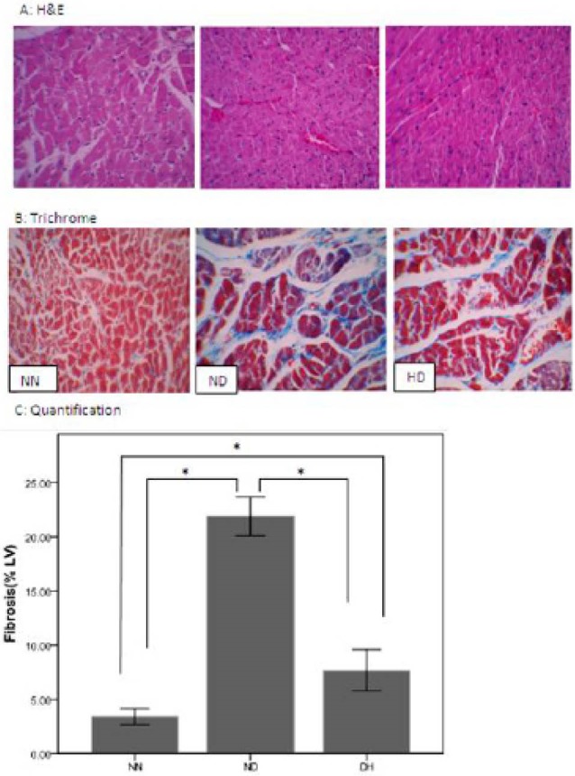
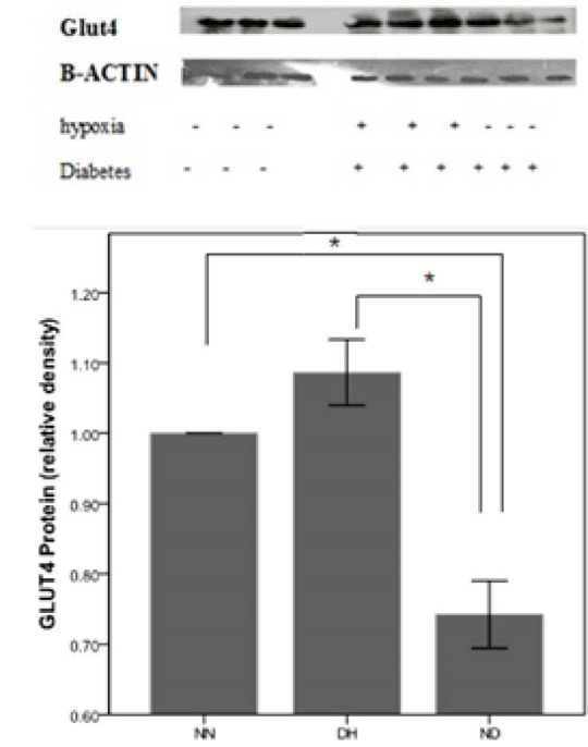
A total number of 30 rats were randomly divided into three groups; 1: Normoxia control group (NN, n = 10). 2: Normoxia diabetic group (ND, n = 10) that took fat diet for 2 weeks then were injected by streptozotocin (37 mg/kg) and 3: Hypoxia diabetic group (HD, n = 10): that were exposed to chronic intermittent hypoxia (CIH) (altitude ≈3400 m, 14% oxygen for 8 weeks). After hypoxia challenge, plasma metabolic parameters including: fasting blood glucose (FBS), triglyceride (TG) and total cholesterol (TC) were measured by colorimetric assay. Cardiac expression of GLUT4 protein and cardiac collagen accumulation were determined in the excised left ventricle by western blotting, and Masson trichrome staining respectively.
Based on resultant data, FBS, TG and TC were significantly (P < 0.05) decreased in HD vs. ND. Homeostasis Model Assessment (HOMA) were also significantly attenuated after exposed to CIH in HD group compared to ND group (P < 0.05). Significant increase in packed cell volume and hemoglobin concentration was observed in HD group compared to ND group (P < 0.05). Comparison of heart wet weight between three groups showed a significant difference (P < 0.05) with lower amount in HD and ND versus NN. Myocardial fibrosis was significantly more pronounced in ND when compared to NN. Eight weeks exposure to hypoxia ameliorated this increase in HD group. Intermittent hypoxia significantly increased GLUT4 protein expression in HD compared to ND group (P < 0.05).
Data suggested that CIH might potentiate to improve glucose homeostasis and cardiac tissue structural damages created in type 2 diabetes (T2D).
慢性间歇性缺氧被认为是心血管健康中的一种预处理状态,可诱导对低氧供应的耐受性。糖尿病性心肌病导致心脏无法有效循环血液,从而防止后续组织损伤。因此,本研究的目的是阐明慢性缺氧暴露对实验性糖尿病性心肌病中心脏纤维化和葡萄糖转运蛋白4(GLUT4)表达的影响。
总共30只大鼠随机分为三组:1. 常氧对照组(NN,n = 10)。2. 常氧糖尿病组(ND,n = 10),给予高脂饮食2周,然后注射链脲佐菌素(37 mg/kg)。3. 缺氧糖尿病组(HD,n = 10):暴露于慢性间歇性缺氧(CIH)(海拔约3400米,14%氧气,持续8周)。缺氧挑战后,通过比色法测量血浆代谢参数,包括空腹血糖(FBS)、甘油三酯(TG)和总胆固醇(TC)。通过蛋白质印迹法和Masson三色染色法分别测定切除的左心室中GLUT4蛋白的心脏表达和心脏胶原积累。
根据所得数据,与ND组相比,HD组的FBS、TG和TC显著降低(P < 0.05)。与ND组相比,HD组暴露于CIH后稳态模型评估(HOMA)也显著减弱(P < 0.05)。与ND组相比,HD组的红细胞压积和血红蛋白浓度显著增加(P < 0.05)。三组之间心脏湿重的比较显示出显著差异(P < 0.05),HD组和ND组低于NN组。与NN组相比,ND组的心肌纤维化明显更明显。缺氧暴露8周改善了HD组的这种增加。与ND组相比,间歇性缺氧显著增加了HD组中GLUT4蛋白的表达(P < 0.05)。
数据表明,CIH可能有助于改善2型糖尿病(T2D)中产生的葡萄糖稳态和心脏组织结构损伤。