Koerfer Justus, Kallendrusch Sonja, Merz Felicitas, Wittekind Christian, Kubick Christoph, Kassahun Woubet T, Schumacher Guido, Moebius Christian, Gaßler Nikolaus, Schopow Nikolas, Geister Daniela, Wiechmann Volker, Weimann Arved, Eckmann Christian, Aigner Achim, Bechmann Ingo, Lordick Florian
Institute for Anatomy, University Medicine Leipzig, Liebigstraße 13, 04103, Leipzig, Germany.
University Cancer Center Leipzig (UCCL), University Medicine Leipzig, Liebigstraße 20, 04103, Leipzig, Germany.
Cancer Med. 2016 Jul;5(7):1444-53. doi: 10.1002/cam4.720. Epub 2016 Apr 12.
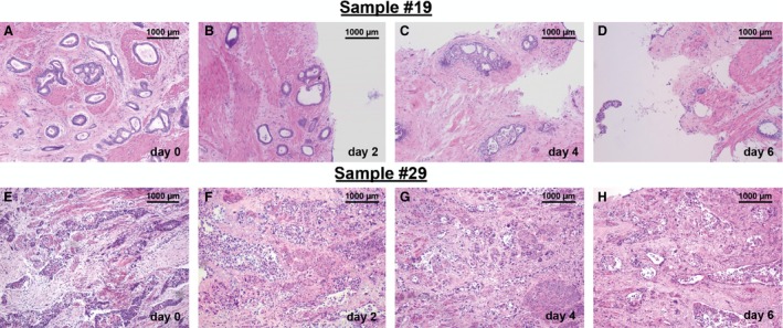
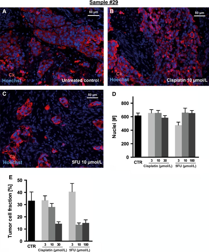
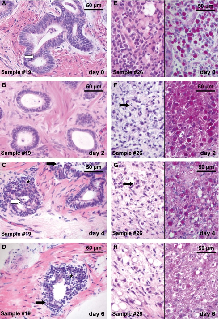
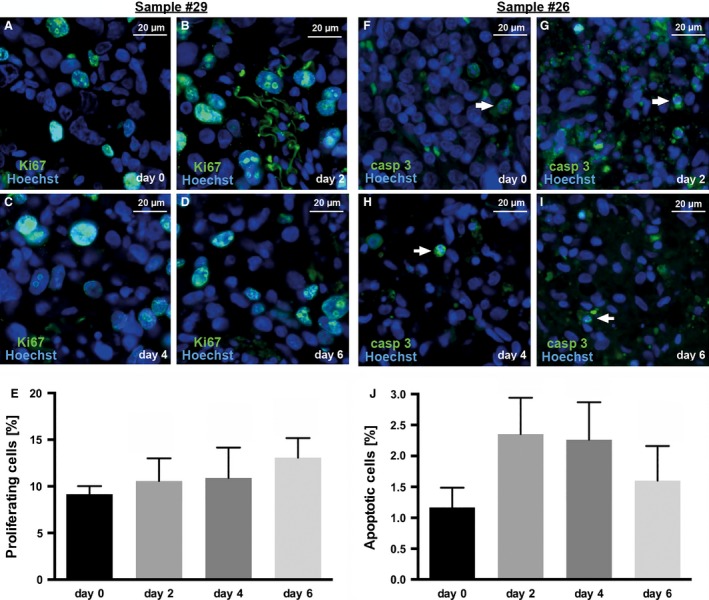
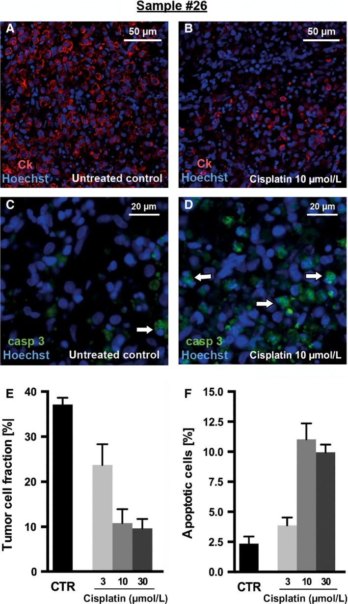
Gastric and esophagogastric junction cancers are heterogeneous and aggressive tumors with an unpredictable response to cytotoxic treatment. New methods allowing for the analysis of drug resistance are needed. Here, we describe a novel technique by which human tumor specimens can be cultured ex vivo, preserving parts of the natural cancer microenvironment. Using a tissue chopper, fresh surgical tissue samples were cut in 400 μm slices and cultivated in 6-well plates for up to 6 days. The slices were processed for routine histopathology and immunohistochemistry. Cytokeratin stains (CK8, AE1/3) were applied for determining tumor cellularity, Ki-67 for proliferation, and cleaved caspase-3 staining for apoptosis. The slices were analyzed under naive conditions and following 2-4 days in vitro exposure to 5-FU and cisplatin. The slice culture technology allowed for a good preservation of tissue morphology and tumor cell integrity during the culture period. After chemotherapy exposure, a loss of tumor cellularity and an increase in apoptosis were observed. Drug sensitivity of the tumors could be assessed. Organotypic slice cultures of gastric and esophagogastric junction cancers were successfully established. Cytotoxic drug effects could be monitored. They may be used to examine mechanisms of drug resistance in human tissue and may provide a unique and powerful ex vivo platform for the prediction of treatment response.
胃癌和食管胃交界癌是异质性侵袭性肿瘤,对细胞毒性治疗的反应难以预测。因此需要新的方法来分析耐药性。在此,我们描述了一种新技术,通过该技术可以对人类肿瘤标本进行体外培养,保留部分天然癌症微环境。使用组织切片机将新鲜手术组织样本切成400μm薄片,并在6孔板中培养长达6天。对切片进行常规组织病理学和免疫组织化学处理。应用细胞角蛋白染色(CK8、AE1/3)确定肿瘤细胞数量,Ki-67检测增殖情况,裂解的半胱天冬酶-3染色检测凋亡情况。在未处理条件下以及体外暴露于5-氟尿嘧啶和顺铂2-4天后对切片进行分析。切片培养技术在培养期间能够很好地保存组织形态和肿瘤细胞完整性。化疗暴露后,观察到肿瘤细胞数量减少且凋亡增加。可以评估肿瘤的药物敏感性。成功建立了胃和食管胃交界癌的器官型切片培养。可以监测细胞毒性药物的作用。它们可用于研究人类组织中的耐药机制,并可能为预测治疗反应提供一个独特而强大的体外平台。