Abreu Maíra Cavallet de, Ponzoni Deise, Langie Renan, Artuzi Felipe Ernesto, Puricelli Edela
Departamento de Cirurgia Oral e Maxilofacial, Faculdade de Odontologia, Universidade Federal do Rio Grande do Sul, Porto Alegre, RS, Brasil.
J Appl Oral Sci. 2016 Apr;24(2):162-70. doi: 10.1590/1678-775720150336.
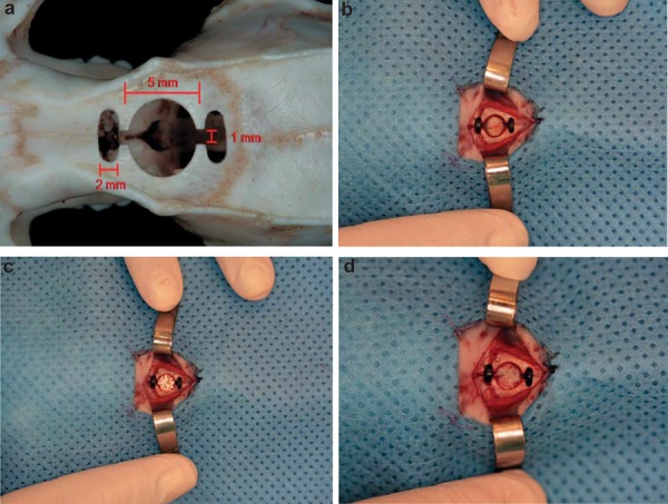
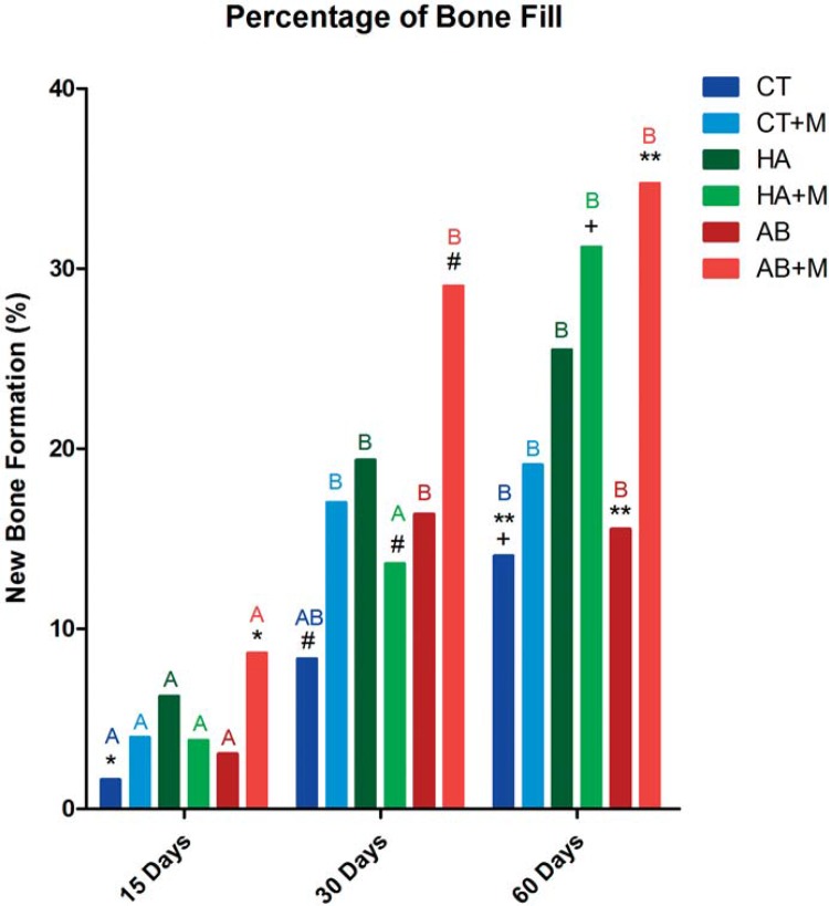
The understanding of bone repair phenomena is a fundamental part of dentistry and maxillofacial surgery. Objective The present study aimed to evaluate the influence of buried magnetic field stimulation on bone repair in rat calvaria after reconstruction with autogenous bone grafts, synthetic powdered hydroxyapatite, or allogeneic cartilage grafts, with or without exposure to magnetic stimulation. Material and Methods Ninety male Wistar rats were divided into 18 groups of five animals each. Critical bone defects were created in the rats' calvaria and immediately reconstructed with autogenous bone, powdered synthetic hydroxyapatite or allogeneic cartilage. Magnetic implants were also placed in half the animals. Rats were euthanized for analysis at 15, 30, and 60 postoperative days. Histomorphometric analyses of the quantity of bone repair were performed at all times. Results These analyses showed significant group by postoperative time interactions (p=0.008). Among the rats subjected to autogenous bone reconstruction, those exposed to magnetic stimulation had higher bone fill percentages than those without magnetic implants. Results also showed that the quality of bone repair remained higher in the former group as compared to the latter at 60 postoperative days. Conclusions After 60 postoperative days, bone repair was greater in the group treated with autogenous bone grafts and exposed to a magnetic field, and bone repair was most pronounced in animals treated with autogenous bone grafts, followed by those treated with powdered synthetic hydroxyapatite and allogeneic cartilage grafts.
对骨修复现象的理解是牙科和颌面外科的一个基本组成部分。目的本研究旨在评估埋藏磁场刺激对自体骨移植、合成羟基磷灰石粉末或同种异体软骨移植重建大鼠颅骨后骨修复的影响,无论是否暴露于磁刺激下。材料与方法将90只雄性Wistar大鼠分为18组,每组5只动物。在大鼠颅骨上制造临界骨缺损,并立即用自体骨、合成羟基磷灰石粉末或同种异体软骨进行重建。半数动物还植入了磁性植入物。在术后15天、30天和60天对大鼠实施安乐死以进行分析。在所有时间点对骨修复量进行组织形态计量学分析。结果这些分析显示组与术后时间存在显著交互作用(p = 0.008)。在接受自体骨重建的大鼠中,暴露于磁刺激的大鼠的骨填充百分比高于未植入磁性植入物的大鼠。结果还表明,术后60天时,前一组的骨修复质量仍高于后一组。结论术后60天时,接受自体骨移植并暴露于磁场的组骨修复效果更好,骨修复在接受自体骨移植的动物中最为明显,其次是接受合成羟基磷灰石粉末和同种异体软骨移植的动物。