Salvador-Culla Borja, Jeong Kyung Jae, Kolovou Paraskevi Evi, Chiang Homer H, Chodosh James, Dohlman Claes H, Kohane Daniel S
Department of Ophthalmology Massachusetts Eye & Ear Infirmary, Harvard Medical School, Boston, MA, USA ; Laboratory for Biomaterials and Drug Delivery, Department of Anesthesiology, Division of Critical Care Medicine, Children's Hospital Boston, Harvard Medical School, Boston, MA, USA ; David H. Koch Institute for Integrative Cancer Research, Massachusetts Institute of Technology, Cambridge, MA, USA.
Department of Chemical Engineering, University of New Hampshire, Durham, NH, USA.
Transl Vis Sci Technol. 2016 Apr 28;5(2):17. doi: 10.1167/tvst.5.2.17. eCollection 2016 Apr.
We tested the feasibility of using titanium to enhance adhesion of the Boston Keratoprosthesis (B-KPro), ultimately to decrease the risk of implant-associated complications.
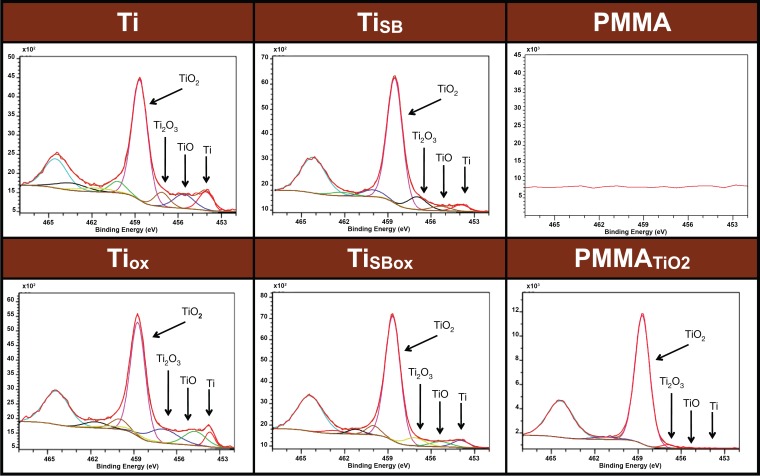
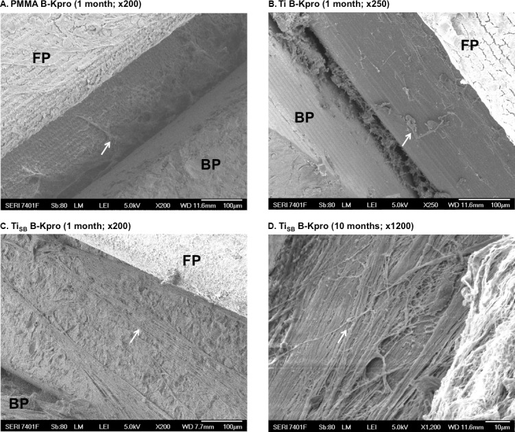
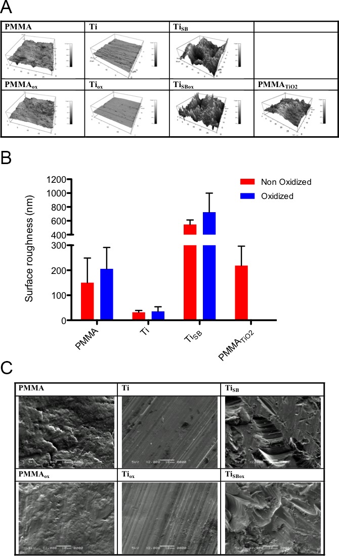
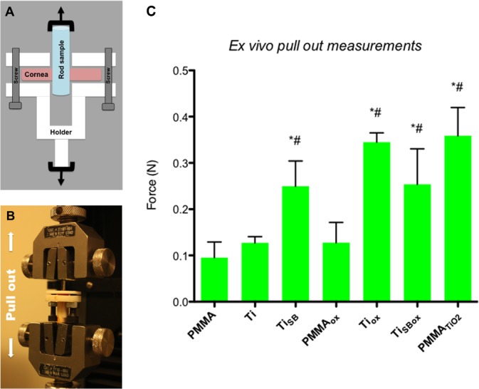
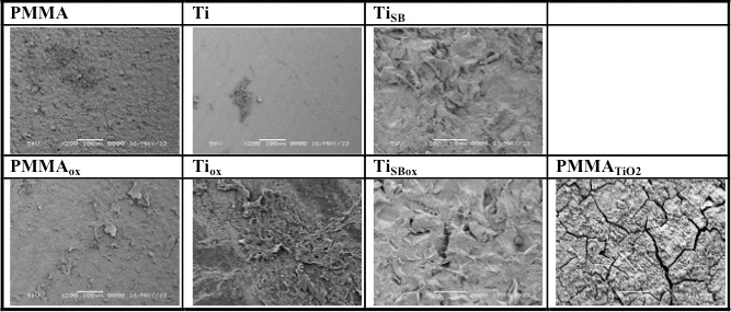
Cylindrical rods were made of poly(methyl methacrylate) (PMMA), PMMA coated with titanium dioxide (TiO) over a layer of polydopamine (PMMA), smooth (Ti) and sandblasted (Ti) titanium, and titanium treated with oxygen plasma (Ti and Ti). Topography and surface chemistry were analyzed by scanning electron microscopy (SEM), atomic force microscopy (AFM), and X-ray photoelectron spectroscopy (XPS). Adhesion force between rods and porcine corneas was measured ex vivo. Titanium sleeves, smooth and sandblasted, were inserted around the stem of the B-KPro and implanted in rabbits. Tissue adhesion to the stem was assessed and compared to an unmodified B-Kpro after 1 month.
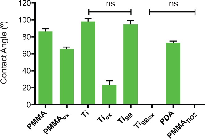
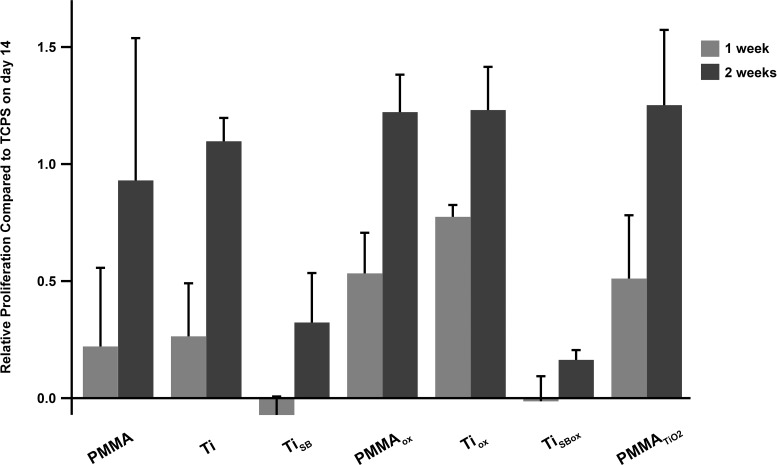
X-ray photoelectron spectroscopy demonstrated successful deposition of TiO on polydopamine-coated PMMA. Oxygen plasma treatment did not change the XPS spectra of titanium rods (Ti and Ti), although it increased their hydrophilicity. The materials did not show cell toxicity. After 14 days of incubation, PMMA, smooth titanium treated with oxygen plasma (Ti), and sandblasted titanium rods (Ti, Ti) showed significantly higher adhesion forces than PMMA ex vivo. In vivo, the use of a Ti sleeve around the stem of the B-KPro induced a significant increase in tissue adhesion compared to a Ti sleeve or bare PMMA.
Sandblasted titanium sleeves greatly enhanced adherence of the B-KPro to the rabbit cornea. This approach may improve adhesion with the donor cornea in humans as well.
This approach may improve adhesion with donor corneas in humans.
我们测试了使用钛来增强波士顿人工角膜(B-KPro)的附着力的可行性,最终降低与植入相关并发症的风险。
圆柱形棒由聚甲基丙烯酸甲酯(PMMA)、在一层聚多巴胺(PMMA)上涂有二氧化钛(TiO)的PMMA、光滑(Ti)和喷砂处理(Ti)的钛以及经氧等离子体处理的钛(Ti和Ti)制成。通过扫描电子显微镜(SEM)、原子力显微镜(AFM)和X射线光电子能谱(XPS)分析其形貌和表面化学性质。在体外测量棒与猪角膜之间的附着力。将光滑和喷砂处理的钛套管插入B-KPro的柄周围并植入兔子体内。1个月后评估组织与柄的附着力,并与未修饰的B-KPro进行比较。
X射线光电子能谱表明TiO成功沉积在聚多巴胺涂层的PMMA上。氧等离子体处理未改变钛棒(Ti和Ti)的XPS光谱,尽管它增加了它们的亲水性。这些材料未显示细胞毒性。孵育14天后,PMMA、经氧等离子体处理的光滑钛(Ti)和喷砂处理的钛棒(Ti、Ti)在体外显示出比PMMA显著更高的附着力。在体内,与钛套管或裸露的PMMA相比,在B-KPro的柄周围使用钛套管可显著增加组织附着力。
喷砂处理的钛套管极大地增强了B-KPro与兔角膜的附着力。这种方法也可能改善其与人类供体角膜的附着力。
这种方法可能改善其与人类供体角膜的附着力。