Vickery Owen N, Machtens Jan-Philipp, Tamburrino Giulia, Seeliger Daniel, Zachariae Ulrich
Computational Biology, School of Life Sciences, University of Dundee, Dow Street, Dundee DD1 5EH, UK; Physics, School of Science and Engineering, University of Dundee, Nethergate, Dundee DD1 4NH, UK.
Forschungszentrum Jülich GmbH, Institute of Complex Systems, Zelluläre Biophysik (ICS-4), Leo-Brandt-Strasse, 52428 Jülich, Germany.
Structure. 2016 Jun 7;24(6):997-1007. doi: 10.1016/j.str.2016.04.007. Epub 2016 May 19.
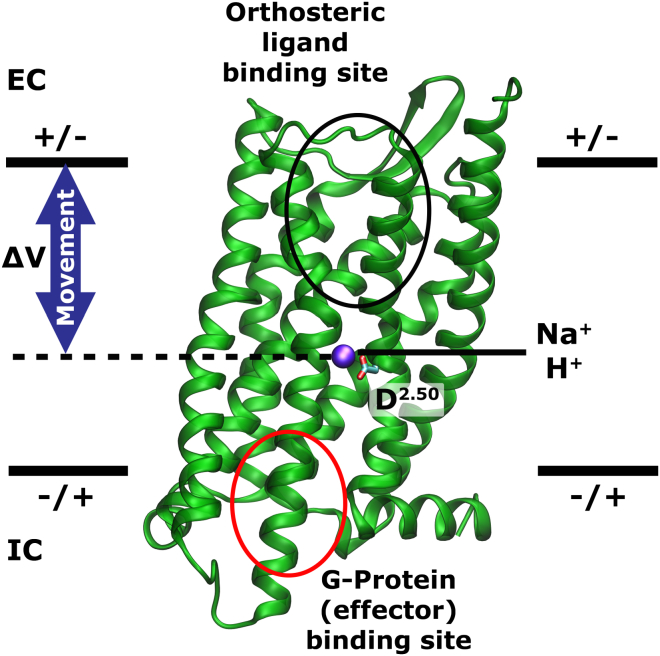
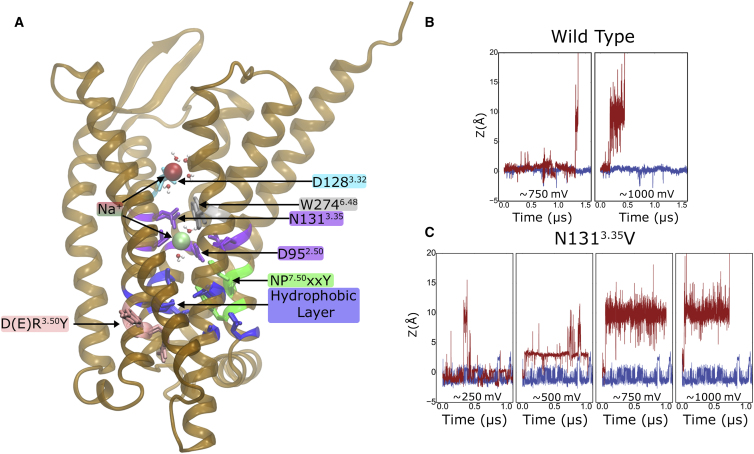
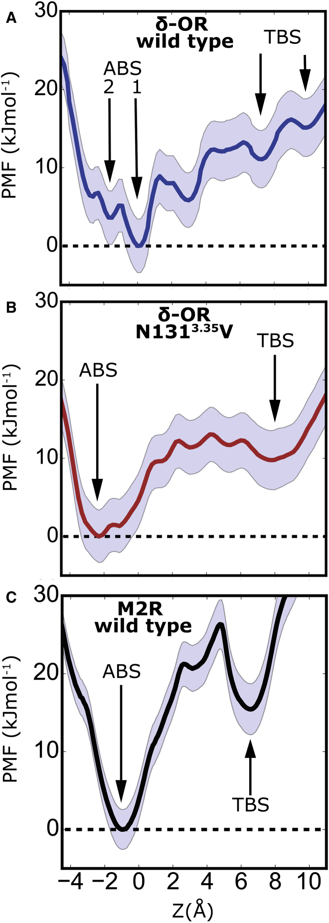
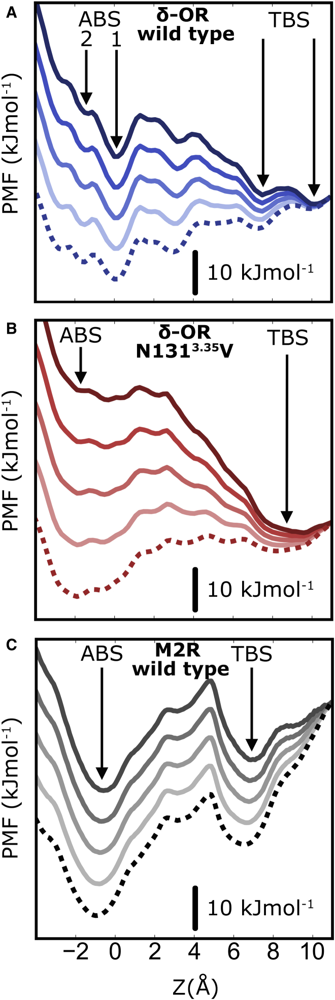
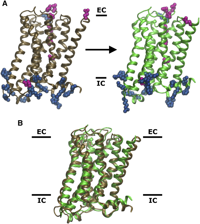
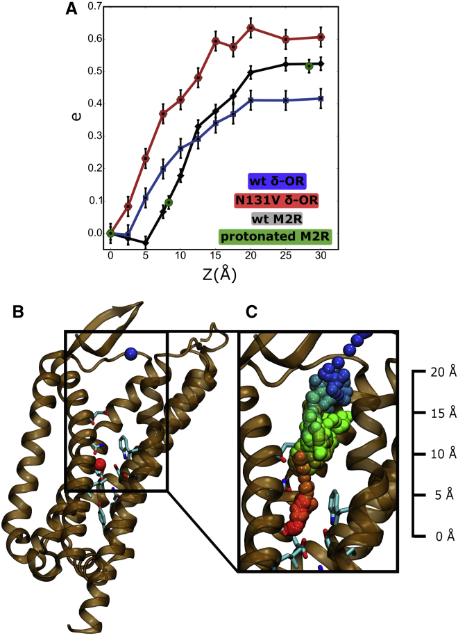
G-protein-coupled receptors (GPCRs) form the largest superfamily of membrane proteins and one-third of all drug targets in humans. A number of recent studies have reported evidence for substantial voltage regulation of GPCRs. However, the structural basis of GPCR voltage sensing has remained enigmatic. Here, we present atomistic simulations on the δ-opioid and M2 muscarinic receptors, which suggest a structural and mechanistic explanation for the observed voltage-induced functional effects. The simulations reveal that the position of an internal Na(+) ion, recently detected to bind to a highly conserved aqueous pocket in receptor crystal structures, strongly responds to voltage changes. The movements give rise to gating charges in excellent agreement with previous experimental recordings. Furthermore, free energy calculations show that these rearrangements of Na(+) can be induced by physiological membrane voltages. Due to its role in receptor function and signal bias, the repositioning of Na(+) has important general implications for signal transduction in GPCRs.
G蛋白偶联受体(GPCRs)构成了膜蛋白中最大的超家族,也是人类所有药物靶点的三分之一。最近的一些研究报告了GPCRs存在显著电压调节的证据。然而,GPCR电压传感的结构基础仍然是个谜。在这里,我们展示了对δ-阿片受体和M2毒蕈碱受体的原子模拟,这为观察到的电压诱导功能效应提供了结构和机制上的解释。模拟结果表明,最近在受体晶体结构中检测到的与一个高度保守的水相口袋结合的内部Na(+)离子的位置,对电压变化有强烈反应。这些移动产生的门控电荷与之前的实验记录非常吻合。此外,自由能计算表明,这些Na(+)的重排可以由生理膜电压诱导。由于其在受体功能和信号偏向中的作用,Na(+)的重新定位对GPCRs中的信号转导具有重要的普遍意义。