• 文献检索
  • 文档翻译
  • 深度研究
  • 学术资讯
  • Suppr Zotero 插件Zotero 插件
  • 邀请有礼
  • 套餐&价格
  • 历史记录
应用&插件
Suppr Zotero 插件Zotero 插件浏览器插件Mac 客户端Windows 客户端微信小程序
定价
高级版会员购买积分包购买API积分包
服务
文献检索文档翻译深度研究API 文档MCP 服务
关于我们
关于 Suppr公司介绍联系我们用户协议隐私条款
关注我们

Suppr 超能文献

核心技术专利:CN118964589B侵权必究
粤ICP备2023148730 号-1Suppr @ 2026

文献检索

告别复杂PubMed语法,用中文像聊天一样搜索,搜遍4000万医学文献。AI智能推荐,让科研检索更轻松。

立即免费搜索

文件翻译

保留排版,准确专业,支持PDF/Word/PPT等文件格式,支持 12+语言互译。

免费翻译文档

深度研究

AI帮你快速写综述,25分钟生成高质量综述,智能提取关键信息,辅助科研写作。

立即免费体验

活性状态 G 蛋白偶联受体中钠离子的细胞内转移

Intracellular Transfer of Na in an Active-State G-Protein-Coupled Receptor.

机构信息

School of Life Sciences, University of Dundee, Dundee DD1 5EH, UK; School of Science and Engineering, University of Dundee, Dundee DD1 4NH, UK.

Department of Biological Sciences, Bridge Institute, University of Southern California, Los Angeles, CA 90089, USA.

出版信息

Structure. 2018 Jan 2;26(1):171-180.e2. doi: 10.1016/j.str.2017.11.013. Epub 2017 Dec 14.

DOI:10.1016/j.str.2017.11.013
PMID:29249607
原文链接:https://pmc.ncbi.nlm.nih.gov/articles/PMC5805466/
Abstract

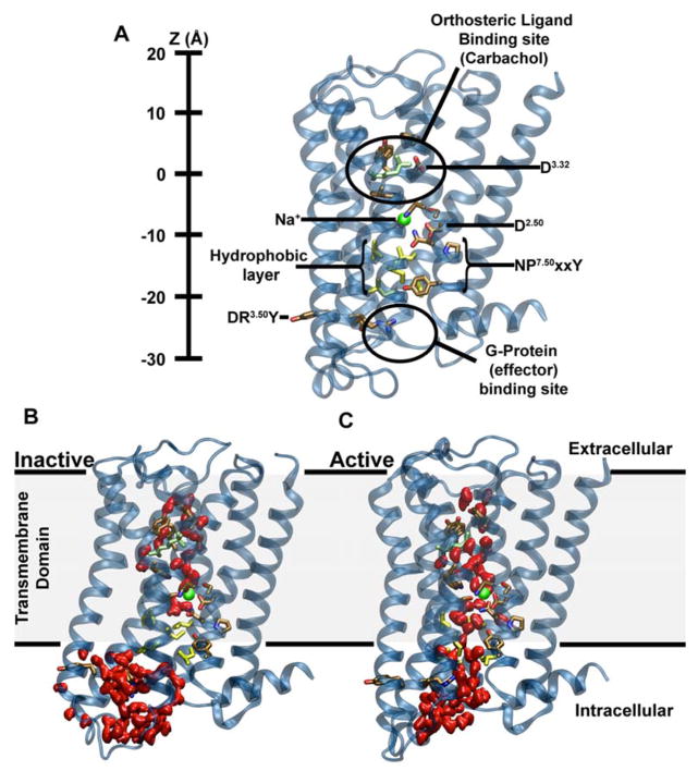
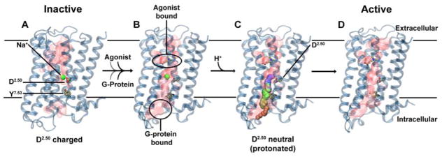
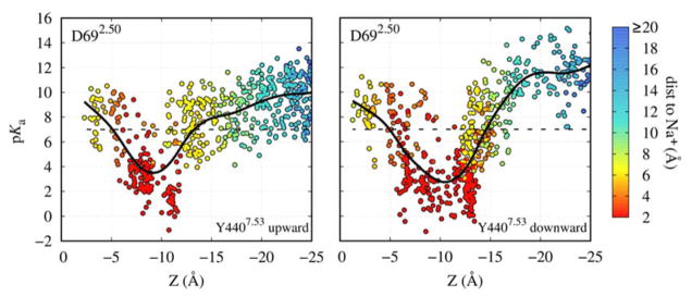
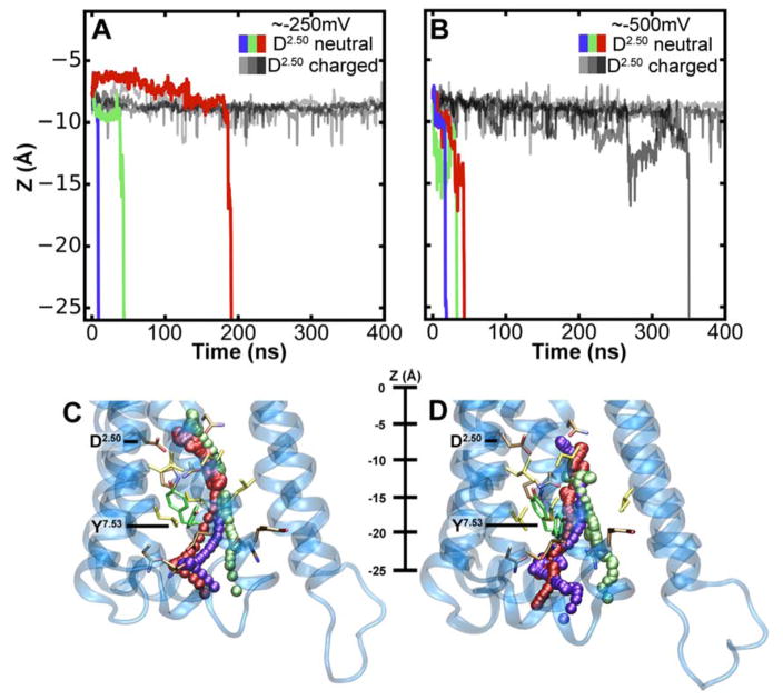
Playing a central role in cell signaling, G-protein-coupled receptors (GPCRs) are the largest superfamily of membrane proteins and form the majority of drug targets in humans. How extracellular agonist binding triggers the activation of GPCRs and associated intracellular effector proteins remains, however, poorly understood. Structural studies have revealed that inactive class A GPCRs harbor a conserved binding site for Na ions in the center of their transmembrane domain, accessible from the extracellular space. Here, we show that the opening of a conserved hydrated channel in the activated state receptors allows the Na ion to egress from its binding site into the cytosol. Coupled with protonation changes, this ion movement occurs without significant energy barriers, and can be driven by physiological transmembrane ion and voltage gradients. We propose that Na ion exchange with the cytosol is a key step in GPCR activation. Further, we hypothesize that this transition locks receptors in long-lived active-state conformations.

摘要

在细胞信号转导中发挥核心作用的 G 蛋白偶联受体(GPCRs)是最大的膜蛋白超家族,也是人类大多数药物靶点的组成部分。然而,细胞外激动剂结合如何触发 GPCR 及其相关细胞内效应蛋白的激活仍然知之甚少。结构研究表明,无活性的 A 类 GPCR 在其跨膜域的中心具有保守的钠离子结合位点,可从细胞外空间进入。在这里,我们表明,在激活状态下,受体中保守的水合通道的打开允许钠离子从其结合位点排出到细胞质中。与质子化变化耦合,这种离子运动不会遇到明显的能量障碍,并且可以由生理跨膜离子和电压梯度驱动。我们提出钠离子与细胞质的交换是 GPCR 激活的关键步骤。此外,我们假设这种转变将受体锁定在长寿命的活性构象中。

https://cdn.ncbi.nlm.nih.gov/pmc/blobs/e019/5805466/761b9b155d2e/nihms939268f6.jpg
https://cdn.ncbi.nlm.nih.gov/pmc/blobs/e019/5805466/d43c8c4b3701/nihms939268f1.jpg
https://cdn.ncbi.nlm.nih.gov/pmc/blobs/e019/5805466/223851e050e9/nihms939268f2.jpg
https://cdn.ncbi.nlm.nih.gov/pmc/blobs/e019/5805466/06f7289ae262/nihms939268f3.jpg
https://cdn.ncbi.nlm.nih.gov/pmc/blobs/e019/5805466/6177c32b1604/nihms939268f4.jpg
https://cdn.ncbi.nlm.nih.gov/pmc/blobs/e019/5805466/023432baf984/nihms939268f5.jpg
https://cdn.ncbi.nlm.nih.gov/pmc/blobs/e019/5805466/761b9b155d2e/nihms939268f6.jpg

相似文献

1
Intracellular Transfer of Na in an Active-State G-Protein-Coupled Receptor.活性状态 G 蛋白偶联受体中钠离子的细胞内转移
Structure. 2018 Jan 2;26(1):171-180.e2. doi: 10.1016/j.str.2017.11.013. Epub 2017 Dec 14.
2
Conformational Complexity and Dynamics in a Muscarinic Receptor Revealed by NMR Spectroscopy.通过 NMR 光谱揭示了毒蕈碱型乙酰胆碱受体的构象复杂性和动态性。
Mol Cell. 2019 Jul 11;75(1):53-65.e7. doi: 10.1016/j.molcel.2019.04.028. Epub 2019 May 15.
3
Graded activation and free energy landscapes of a muscarinic G-protein-coupled receptor.毒蕈碱型 G 蛋白偶联受体的分级激活与自由能景观
Proc Natl Acad Sci U S A. 2016 Oct 25;113(43):12162-12167. doi: 10.1073/pnas.1614538113. Epub 2016 Oct 10.
4
Structural Mechanisms of Voltage Sensing in G Protein-Coupled Receptors.G蛋白偶联受体中电压感应的结构机制
Structure. 2016 Jun 7;24(6):997-1007. doi: 10.1016/j.str.2016.04.007. Epub 2016 May 19.
5
Protonation state of the selectivity filter of bacterial voltage-gated sodium channels is modulated by ions.细菌电压门控钠离子通道的选择性过滤器的质子化状态受离子调节。
Proteins. 2020 Mar;88(3):527-539. doi: 10.1002/prot.25831. Epub 2019 Nov 12.
6
Sodium ions allosterically modulate the M2 muscarinic receptor.钠离子变构调节 M2 毒蕈碱型乙酰胆碱受体。
Sci Rep. 2020 Jul 7;10(1):11177. doi: 10.1038/s41598-020-68133-9.
7
Conformational changes in the M2 muscarinic receptor induced by membrane voltage and agonist binding.M2 毒蕈碱型乙酰胆碱受体构象变化:由膜电压和激动剂结合诱导。
J Physiol. 2011 Apr 1;589(Pt 7):1741-53. doi: 10.1113/jphysiol.2010.204107. Epub 2011 Jan 31.
8
ASIC and ENaC type sodium channels: conformational states and the structures of the ion selectivity filters.ASIC 和 ENaC 型钠离子通道:构象状态和离子选择性过滤器的结构。
FEBS J. 2017 Feb;284(4):525-545. doi: 10.1111/febs.13840. Epub 2016 Sep 15.
9
Universality of the Sodium Ion Binding Mechanism in Class A G-Protein-Coupled Receptors.A 类 G 蛋白偶联受体钠离子结合机制的普遍性。
Angew Chem Int Ed Engl. 2018 Mar 12;57(12):3048-3053. doi: 10.1002/anie.201708889. Epub 2018 Feb 22.
10
Activation and dynamic network of the M2 muscarinic receptor.M2 毒蕈碱型乙酰胆碱受体的激活和动态网络。
Proc Natl Acad Sci U S A. 2013 Jul 2;110(27):10982-7. doi: 10.1073/pnas.1309755110. Epub 2013 Jun 18.

引用本文的文献

1
Integrated bulk RNA and single-cell analysis with experimental validation reveal oxidative stress-related diagnostic biomarkers for osteoporosis.整合批量RNA和单细胞分析并进行实验验证,揭示了骨质疏松症的氧化应激相关诊断生物标志物。
PLoS One. 2025 Apr 29;20(4):e0322326. doi: 10.1371/journal.pone.0322326. eCollection 2025.
2
The structural basis of the G protein-coupled receptor and ion channel axis.G蛋白偶联受体与离子通道轴的结构基础。
Curr Res Struct Biol. 2025 Feb 18;9:100165. doi: 10.1016/j.crstbi.2025.100165. eCollection 2025 Jun.
3
Structure-guided design of partial agonists at an opioid receptor.

本文引用的文献

1
Improving efficiency of large time-scale molecular dynamics simulations of hydrogen-rich systems.提高富氢体系大时间尺度分子动力学模拟的效率。
J Comput Chem. 1999 Jun;20(8):786-798. doi: 10.1002/(SICI)1096-987X(199906)20:8<786::AID-JCC5>3.0.CO;2-B.
2
Gating Charge Calculations by Computational Electrophysiology Simulations.通过计算电生理模拟进行门控电荷计算
Biophys J. 2017 Apr 11;112(7):1396-1405. doi: 10.1016/j.bpj.2017.02.016.
3
Mechanistic insights into GPCR-G protein interactions.对G蛋白偶联受体(GPCR)与G蛋白相互作用的机制性见解。
阿片受体部分激动剂的结构导向设计。
Nat Commun. 2025 Mar 13;16(1):2518. doi: 10.1038/s41467-025-57734-5.
4
Computational design of highly signalling-active membrane receptors through solvent-mediated allosteric networks.通过溶剂介导的变构网络进行高信号活性膜受体的计算设计
Nat Chem. 2025 Mar;17(3):429-438. doi: 10.1038/s41557-024-01719-2. Epub 2025 Jan 23.
5
Systematic analysis of biomolecular conformational ensembles with PENSA.使用PENSA对生物分子构象集合进行系统分析。
J Chem Phys. 2025 Jan 7;162(1). doi: 10.1063/5.0235544.
6
Mechanical stress and anionic lipids synergistically stabilize an atypical structure of the angiotensin II type 1 receptor (AT1).机械应力和阴离子脂质协同稳定血管紧张素 II 型 1 型受体 (AT1) 的非典型结构。
PLoS Comput Biol. 2024 Nov 13;20(11):e1012559. doi: 10.1371/journal.pcbi.1012559. eCollection 2024 Nov.
7
p Calculations of GPCRs: Understanding Protonation States in Receptor Activation.GPCRs 的计算:理解受体激活中的质子化状态。
J Chem Inf Model. 2024 Sep 9;64(17):6850-6856. doi: 10.1021/acs.jcim.4c01125. Epub 2024 Aug 16.
8
Protons taken hostage: Dynamic H-bond networks of the pH-sensing GPR68.被“挟持”的质子:pH 传感受体 GPR68 的动态氢键网络
Comput Struct Biotechnol J. 2023 Sep 2;21:4370-4384. doi: 10.1016/j.csbj.2023.08.034. eCollection 2023.
9
The World of GPCR dimers - Mapping dopamine receptor D homodimers in different activation states and configuration arrangements.GPCR二聚体的世界——绘制处于不同激活状态和构象排列的多巴胺受体D同型二聚体图谱。
Comput Struct Biotechnol J. 2023 Sep 3;21:4336-4353. doi: 10.1016/j.csbj.2023.08.032. eCollection 2023.
10
Three-Dimensional Structural Insights Have Revealed the Distinct Binding Interactions of Agonists, Partial Agonists, and Antagonists with the µ Opioid Receptor.三维结构见解揭示了激动剂、部分激动剂和拮抗剂与μ阿片受体的独特结合相互作用。
Int J Mol Sci. 2023 Apr 11;24(8):7042. doi: 10.3390/ijms24087042.
Curr Opin Struct Biol. 2016 Dec;41:247-254. doi: 10.1016/j.sbi.2016.11.005. Epub 2016 Nov 18.
4
GPCR-G Protein-β-Arrestin Super-Complex Mediates Sustained G Protein Signaling.G蛋白偶联受体-G蛋白-β-抑制蛋白超级复合物介导持续的G蛋白信号传导。
Cell. 2016 Aug 11;166(4):907-919. doi: 10.1016/j.cell.2016.07.004. Epub 2016 Aug 4.
5
Membrane potentials regulating GPCRs: insights from experiments and molecular dynamics simulations.调节G蛋白偶联受体的膜电位:来自实验和分子动力学模拟的见解
Curr Opin Pharmacol. 2016 Oct;30:44-50. doi: 10.1016/j.coph.2016.06.011. Epub 2016 Jul 27.
6
Guiding lead optimization with GPCR structure modeling and molecular dynamics.利用GPCR结构建模和分子动力学指导先导物优化。
Curr Opin Pharmacol. 2016 Oct;30:14-21. doi: 10.1016/j.coph.2016.06.004. Epub 2016 Jul 13.
7
Allosteric coupling from G protein to the agonist-binding pocket in GPCRs.G蛋白偶联受体中从G蛋白到激动剂结合口袋的变构偶联。
Nature. 2016 Jul 7;535(7610):182-6. doi: 10.1038/nature18324. Epub 2016 Jun 29.
8
Mechanistic Studies on the Stereoselectivity of the Serotonin 5-HT1A Receptor.关于血清素 5-HT1A 受体立体选择性的机制研究。
Angew Chem Int Ed Engl. 2016 Jul 18;55(30):8661-5. doi: 10.1002/anie.201603766. Epub 2016 May 31.
9
Structural Mechanisms of Voltage Sensing in G Protein-Coupled Receptors.G蛋白偶联受体中电压感应的结构机制
Structure. 2016 Jun 7;24(6):997-1007. doi: 10.1016/j.str.2016.04.007. Epub 2016 May 19.
10
The agonist-specific voltage dependence of M2 muscarinic receptors modulates the deactivation of the acetylcholine-gated K(+) current (I KACh).M2毒蕈碱受体的激动剂特异性电压依赖性调节乙酰胆碱门控钾电流(IKACh)的失活。
Pflugers Arch. 2016 Jul;468(7):1207-1214. doi: 10.1007/s00424-016-1812-y. Epub 2016 Mar 29.