Viral Diseases Division, National Microbiology Laboratory, Public Health Agency of Canada, Winnipeg, Manitoba, Canada.
Department of Medical Microbiology, University of Manitoba, Winnipeg, Manitoba, Canada.
Sci Rep. 2016 May 23;6:26516. doi: 10.1038/srep26516.
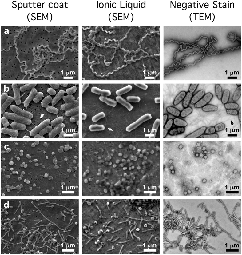
Despite being an excellent tool for investigating ultrastructure, scanning electron microscopy (SEM) is less frequently used than transmission electron microscopy for microbes such as viruses or bacteria. Here we describe rapid methods that allow SEM imaging of fully hydrated, unfixed microbes without using conventional sample preparation methods. We demonstrate improved ultrastructural preservation, with greatly reduced dehydration and shrinkage, for specimens including bacteria and viruses such as Ebola virus using infiltration with ionic liquid on conducting filter substrates for SEM.
尽管扫描电子显微镜(SEM)是一种研究超微结构的优秀工具,但对于病毒或细菌等微生物,其使用频率低于透射电子显微镜。本文介绍了无需采用传统样品制备方法,即可对完全水合、未经固定的微生物进行 SEM 成像的快速方法。我们通过在导电滤膜基底上用离子液体对包括细菌和埃博拉病毒在内的样本进行渗透,实现了更好的超微结构保存,大大减少了脱水和收缩。