Yang Helen H, St-Pierre François, Sun Xulu, Ding Xiaozhe, Lin Michael Z, Clandinin Thomas R
Department of Neurobiology, Stanford University, Stanford, CA 94305, USA.
Department of Bioengineering, Stanford University, Stanford, CA 94305, USA.
Cell. 2016 Jun 30;166(1):245-57. doi: 10.1016/j.cell.2016.05.031. Epub 2016 Jun 2.
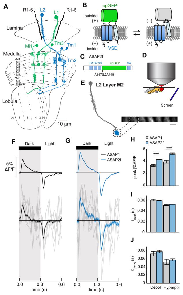
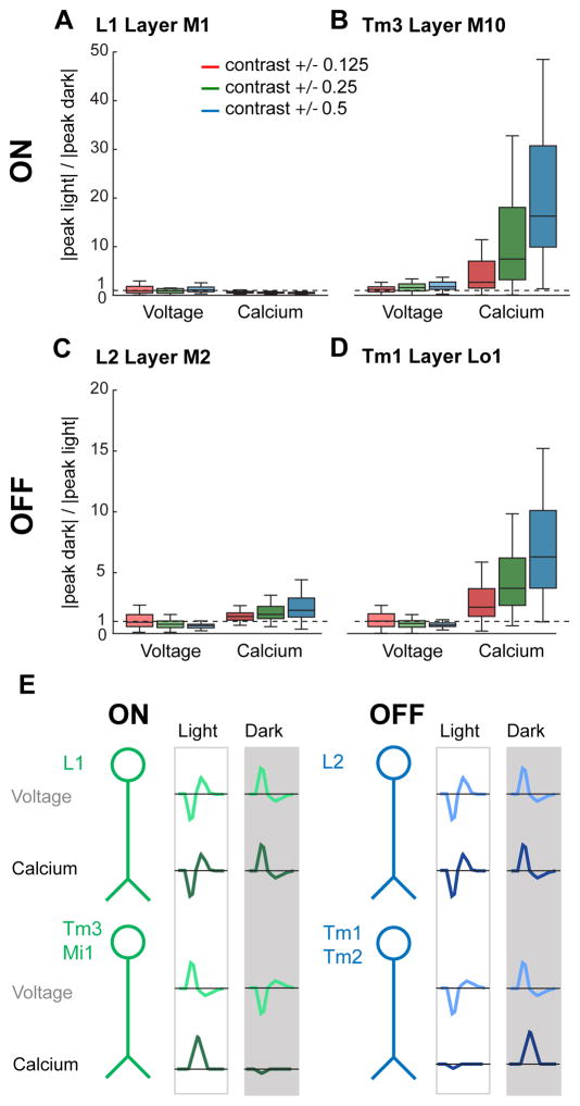
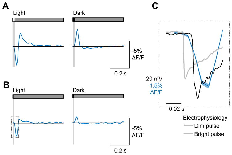
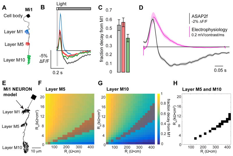
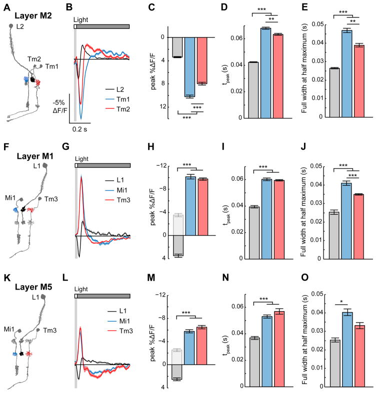
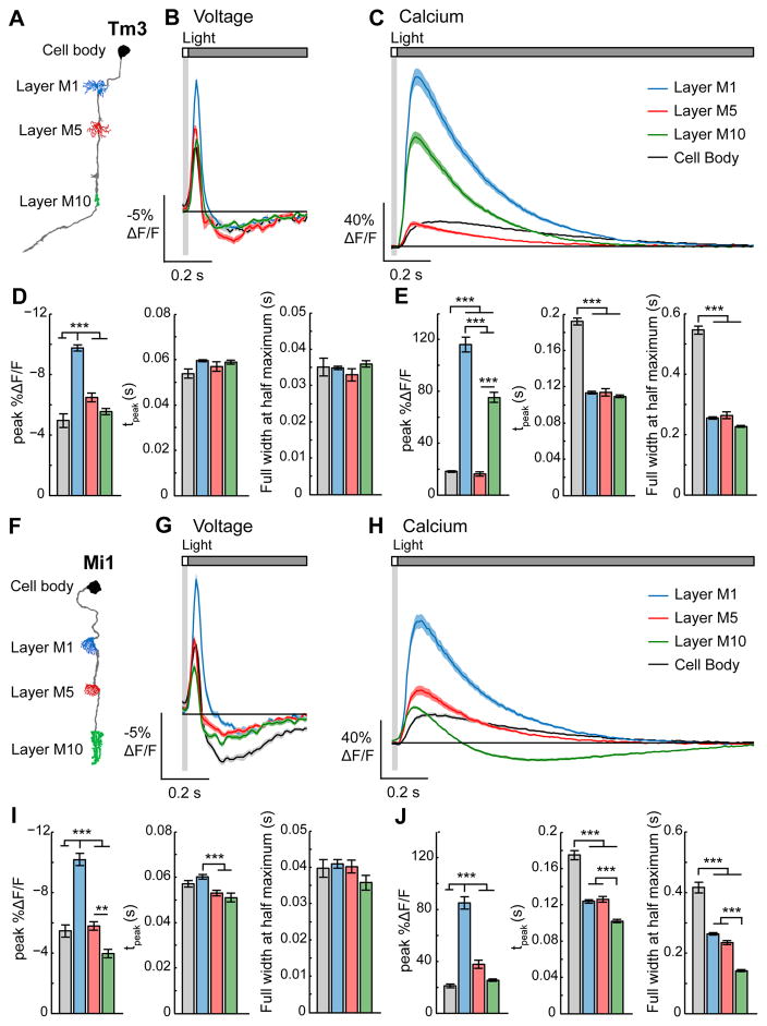
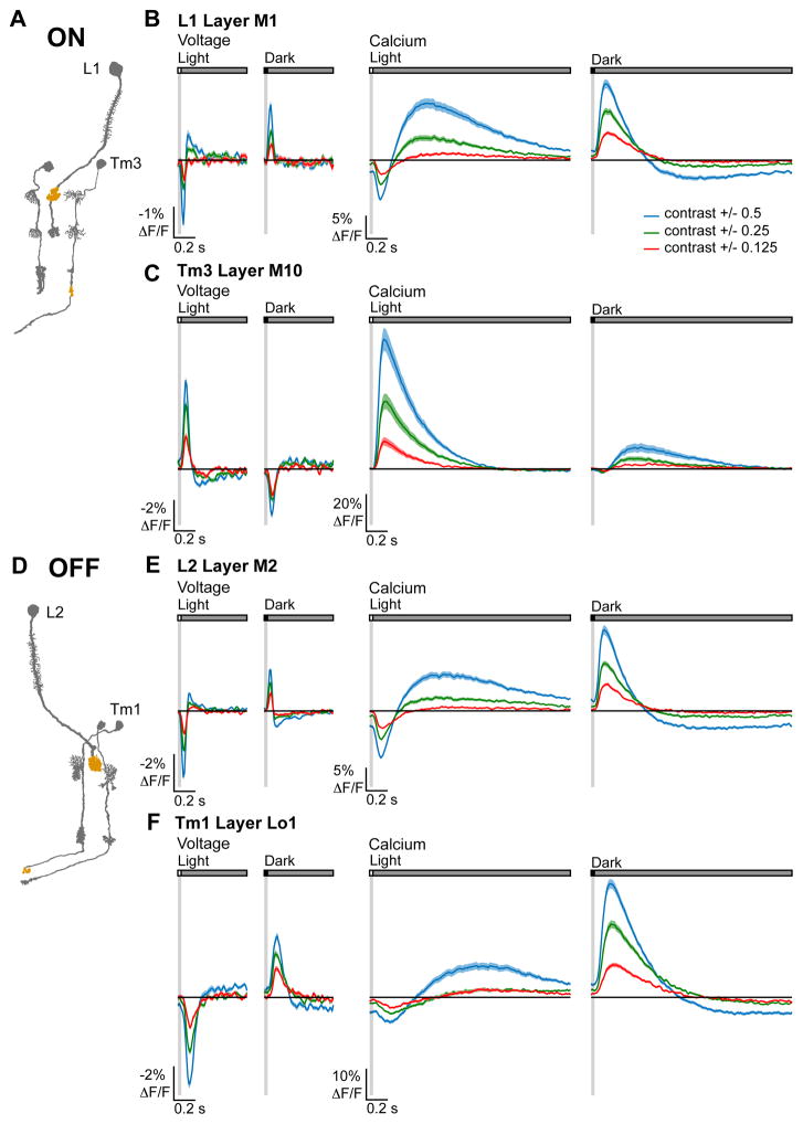
A mechanistic understanding of neural computation requires determining how information is processed as it passes through neurons and across synapses. However, it has been challenging to measure membrane potential changes in axons and dendrites in vivo. We use in vivo, two-photon imaging of novel genetically encoded voltage indicators, as well as calcium imaging, to measure sensory stimulus-evoked signals in the Drosophila visual system with subcellular resolution. Across synapses, we find major transformations in the kinetics, amplitude, and sign of voltage responses to light. We also describe distinct relationships between voltage and calcium signals in different neuronal compartments, a substrate for local computation. Finally, we demonstrate that ON and OFF selectivity, a key feature of visual processing across species, emerges through the transformation of membrane potential into intracellular calcium concentration. By imaging voltage and calcium signals to map information flow with subcellular resolution, we illuminate where and how critical computations arise.
对神经计算的机制性理解需要确定信息在通过神经元和跨越突触时是如何被处理的。然而,在体内测量轴突和树突中的膜电位变化一直具有挑战性。我们利用新型基因编码电压指示剂的体内双光子成像以及钙成像,以亚细胞分辨率测量果蝇视觉系统中感觉刺激诱发的信号。在整个突触中,我们发现光诱发的电压反应在动力学、幅度和符号上有重大转变。我们还描述了不同神经元区室中电压信号和钙信号之间的独特关系,这是局部计算的基础。最后,我们证明,开和关选择性作为跨物种视觉处理的一个关键特征,是通过膜电位向细胞内钙浓度的转变而出现的。通过对电压和钙信号进行成像,以亚细胞分辨率绘制信息流,我们阐明了关键计算发生的位置和方式。