Hao Xiaoli, Yi Changxian, Wang Yuqin, Li Jin, Huang Fang, He Liwen, Chi Wei
State Key Laboratory of Ophthalmology,Zhongshan Ophthalmic Center,Sun Yat-Sen University,Guangzhou, China; Department of Ophthalmology, Institute of Surgery Research, Daping Hospital, Third Military Medical University, Chongqing, China.
State Key Laboratory of Ophthalmology,Zhongshan Ophthalmic Center,Sun Yat-Sen University,Guangzhou, China.
Mol Vis. 2016 Jun 2;22:563-74. eCollection 2016.
Endophthalmitis is mediated by inflammatory cytokines. We employed a quantitative antibody array, which profiles protein expression and function in a high-throughput manner, to identify inflammatory mediators in the infectious aqueous and vitreous humor from patients with endophthalmitis.
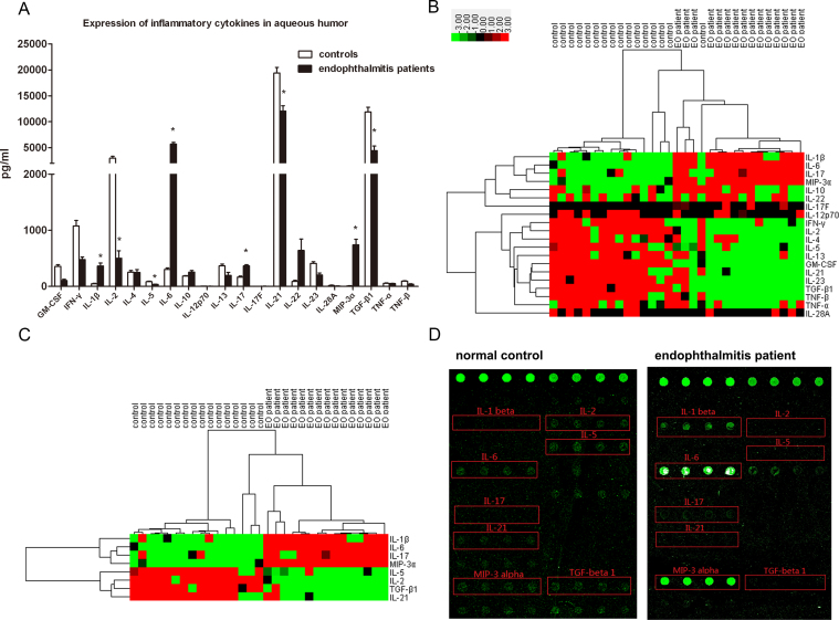
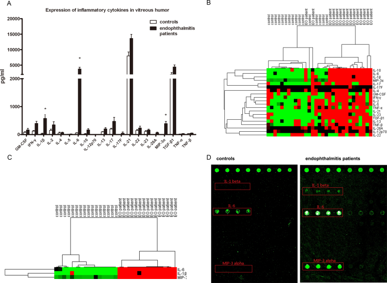
In this prospective study, aqueous humor (AH) and vitreous humor (VH) samples were obtained from 30 patients with endophthalmitis and were collected during anterior chamber paracentesis and vitrectomy. Control samples were obtained from 32 healthy donors. We examined the expression of 20 inflammatory mediators in AH and VH using a quantitative antibody protein array. Hierarchical cluster analysis based on the expression of the quantified cytokines was applied to identify the specificity of endophthalmitis disease. Validation analysis using enzyme-linked immunosorbent assay (ELISA) was performed to confirm the expression of the cytokines identified in the AH and VH samples.
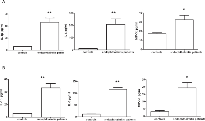
Our results demonstrated elevated expression of interleukin (IL)-1β, IL-6, and macrophage inflammatory protein (MIP)-3α in AH or VH from patients with endophthalmitis. The concentration of IL-17 was upregulated in AH from the patients. The expression of IL-2, IL-5, IL-21, and transforming growth factor (TGF)-β1 was downregulated in AH from the patients. The cluster analysis demonstrated that the cytokine profile expression in AH or VH significantly differed between the patients with endophthalmitis and the healthy controls. Confirmation with ELISA validated the increase in IL-1β, IL-6, and MIP-3α in the AH and VH samples from the patients with endophthalmitis.
Increased expression of IL-1β, IL-6, IL-17, and MIP-3α and decreased expression of IL-2, IL-5, IL-21, and TGF-β in the AH and VH suggests an abnormal cytokine profile in patients with endophthalmitis. Knowledge of this will aid in the diagnosis of infectious endophthalmitis.
眼内炎由炎性细胞因子介导。我们采用定量抗体芯片,以高通量方式分析蛋白质表达及功能,以鉴定眼内炎患者感染性房水和玻璃体内的炎性介质。
在这项前瞻性研究中,从30例眼内炎患者的前房穿刺术和玻璃体切割术中获取房水(AH)和玻璃体(VH)样本。对照样本来自32名健康供体。我们使用定量抗体蛋白芯片检测AH和VH中20种炎性介质的表达。基于定量细胞因子的表达进行层次聚类分析,以确定眼内炎疾病的特异性。采用酶联免疫吸附测定(ELISA)进行验证分析,以确认AH和VH样本中鉴定出的细胞因子的表达。
我们的结果表明,眼内炎患者的AH或VH中白细胞介素(IL)-1β、IL-6和巨噬细胞炎性蛋白(MIP)-3α表达升高。患者AH中IL-17浓度上调。患者AH中IL-2、IL-5、IL-21和转化生长因子(TGF)-β1表达下调。聚类分析表明,眼内炎患者与健康对照者的AH或VH中细胞因子谱表达存在显著差异。ELISA验证证实了眼内炎患者AH和VH样本中IL-1β、IL-6和MIP-3α的增加。
AH和VH中IL-1β、IL-6、IL-17和MIP-3α表达增加,IL-2、IL-5、IL-21和TGF-β表达降低,提示眼内炎患者细胞因子谱异常。了解这一点将有助于感染性眼内炎的诊断。