Duncan James M, Nahas Samuel, Akhtar Kashif, Daurka Jasvinder
Kings College Hospital, London.
Imperial College Hospitals NHS Trust, London.
J Orthop Case Rep. 2015 Jan-Mar;5(1):23-5. doi: 10.13107/jocr.2250-0685.247.
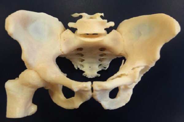
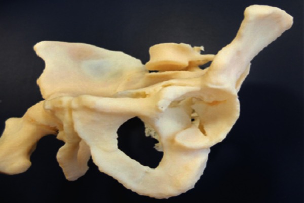
Surgical management of acetabular fractures is often highly complex, and a successful outcome depends upon an appreciation of the fracture pattern and the most appropriate approach to reduce and hold it. Currently, computed tomography (CT) images are used in conjunction with plain x-rays to identify the main fracture components and their spatial relationship to one another, and as such surgeons still have to make decisions based upon their ability to visualise the fracture from the images available. 3D printers have now become widely available and inexpensive, and can be used to rapidly produce life-size models based on CT scans of an individual patient. The availability of patient specific, accurate and detailed models of complex acetabular fractures can aid planning of surgical management on a patient specific basis.
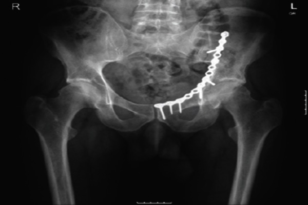
This report describes the use of a 3D printer to create a life-size model reconstruction of the pelvis of a 48 year old male patient who sustained a left sided associated both column acetabular fracture following a motorbike accident in the Sahara Desert. The model allowed visualisation of the multiple fracture fragments and their relative displacements. The tactile feedback allowed assessment of the different fracture fragments. The relative displacement of the quadrilateral plate and posterior column fragments could be assessed and the surgeon felt that these would be amenable to reduction from an ilioinguinal approach. An anatomic reduction was achieved and was held with the application of a pelvic brim plate with 2 screws lagging the posterior column/quadrilateral plate fragment.
There are previous examples of 3D models being used in orthopaedic surgery through the use of rapid prototyping, however this method is usually expensive and time consuming. Advances in 3D printer technology offer surgeons a number of advantages when treating these complex fractures. With the ever-increasing economy, ease of use and speed of additive processing, the possible applications of this technology within orthopaedic surgery are numerous. Given the possible applications of this technology, and its ever increasing availability, we feel that its use can only improve patient outcomes and so should be explored further for use in orthopaedic surgery.
髋臼骨折的手术治疗通常极为复杂,成功的治疗结果取决于对骨折类型的认识以及采用最合适的方法进行复位和固定。目前,计算机断层扫描(CT)图像与普通X线片联合使用,以识别主要骨折块及其相互间的空间关系,因此外科医生仍需凭借从现有图像中可视化骨折的能力来做出决策。3D打印机现已广泛普及且价格低廉,可用于根据个体患者的CT扫描快速制作实物大小的模型。复杂髋臼骨折的患者特异性、准确且详细的模型,有助于基于患者个体情况进行手术治疗规划。
本报告描述了使用3D打印机为一名48岁男性患者创建骨盆实物大小的模型重建,该患者在撒哈拉沙漠遭遇摩托车事故后,左侧发生双柱髋臼骨折。该模型能够可视化多个骨折碎片及其相对位移。触觉反馈有助于评估不同的骨折碎片。可以评估四边形板和后柱碎片的相对位移,外科医生认为这些骨折块适合通过髂腹股沟入路进行复位。通过应用骨盆缘钢板并使用2枚螺钉固定后柱/四边形板碎片,实现了解剖复位。
以往有通过快速成型技术在骨科手术中使用3D模型的例子,但这种方法通常昂贵且耗时。3D打印机技术的进步为外科医生治疗这些复杂骨折提供了诸多优势。随着经济性、易用性和增材加工速度的不断提高,该技术在骨科手术中的潜在应用众多。鉴于该技术的潜在应用及其日益普及,我们认为其应用只会改善患者预后,因此应在骨科手术中进一步探索其用途。