• 文献检索
  • 文档翻译
  • 深度研究
  • 学术资讯
  • Suppr Zotero 插件Zotero 插件
  • 邀请有礼
  • 套餐&价格
  • 历史记录
应用&插件
Suppr Zotero 插件Zotero 插件浏览器插件Mac 客户端Windows 客户端微信小程序
定价
高级版会员购买积分包购买API积分包
服务
文献检索文档翻译深度研究API 文档MCP 服务
关于我们
关于 Suppr公司介绍联系我们用户协议隐私条款
关注我们

Suppr 超能文献

核心技术专利:CN118964589B侵权必究
粤ICP备2023148730 号-1Suppr @ 2026

文献检索

告别复杂PubMed语法,用中文像聊天一样搜索,搜遍4000万医学文献。AI智能推荐,让科研检索更轻松。

立即免费搜索

文件翻译

保留排版,准确专业,支持PDF/Word/PPT等文件格式,支持 12+语言互译。

免费翻译文档

深度研究

AI帮你快速写综述,25分钟生成高质量综述,智能提取关键信息,辅助科研写作。

立即免费体验

游离的主链羰基介导视紫红质激活。

Free backbone carbonyls mediate rhodopsin activation.

作者信息

Kimata Naoki, Pope Andreyah, Sanchez-Reyes Omar B, Eilers Markus, Opefi Chikwado A, Ziliox Martine, Reeves Philip J, Smith Steven O

机构信息

Department of Biochemistry and Cell Biology, Stony Brook University, Stony Brook, New York, USA.

School of Biological Sciences, University of Essex, Essex, UK.

出版信息

Nat Struct Mol Biol. 2016 Aug;23(8):738-43. doi: 10.1038/nsmb.3257. Epub 2016 Jul 4.

DOI:10.1038/nsmb.3257
PMID:27376589
原文链接:https://pmc.ncbi.nlm.nih.gov/articles/PMC4972713/
Abstract

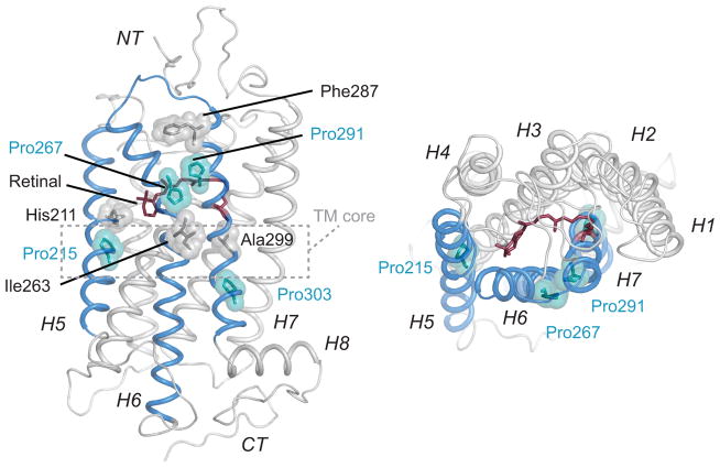
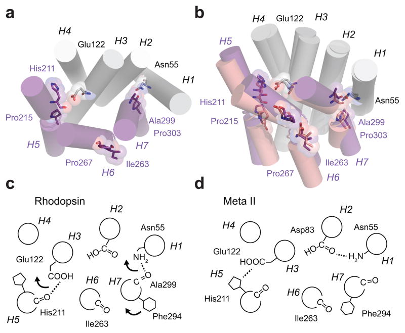
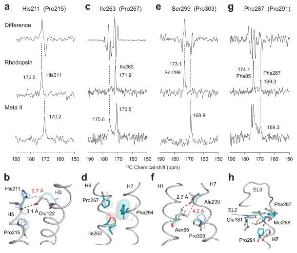
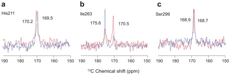
Conserved prolines in the transmembrane helices of G-protein-coupled receptors (GPCRs) are often considered to function as hinges that divide the helix into two segments capable of independent motion. Depending on their potential to hydrogen-bond, the free C=O groups associated with these prolines can facilitate conformational flexibility, conformational switching or stabilization of the receptor structure. To address the role of conserved prolines in family A GPCRs through solid-state NMR spectroscopy, we focus on bovine rhodopsin, a GPCR in the visual receptor subfamily. The free backbone C=O groups on helices H5 and H7 stabilize the inactive rhodopsin structure through hydrogen-bonds to residues on adjacent helices. In response to light-induced isomerization of the retinal chromophore, hydrogen-bonding interactions involving these C=O groups are released, thus facilitating repacking of H5 and H7 onto the transmembrane core of the receptor. These results provide insights into the multiple structural and functional roles of prolines in membrane proteins.

摘要

G蛋白偶联受体(GPCRs)跨膜螺旋中的保守脯氨酸通常被认为起到铰链的作用,将螺旋分为能够独立运动的两个部分。根据其形成氢键的潜力,与这些脯氨酸相关的游离C=O基团可以促进受体结构的构象灵活性、构象转换或稳定。为了通过固态核磁共振光谱研究保守脯氨酸在A类GPCRs中的作用,我们聚焦于牛视紫红质,它是视觉受体亚家族中的一种GPCR。H5和H7螺旋上的游离主链C=O基团通过与相邻螺旋上的残基形成氢键来稳定无活性的视紫红质结构。响应于视黄醛发色团的光诱导异构化,涉及这些C=O基团的氢键相互作用被释放,从而促进H5和H7重新排列到受体的跨膜核心上。这些结果为脯氨酸在膜蛋白中的多种结构和功能作用提供了见解。

https://cdn.ncbi.nlm.nih.gov/pmc/blobs/8098/4972713/a6c2099d985d/nihms794593f4.jpg
https://cdn.ncbi.nlm.nih.gov/pmc/blobs/8098/4972713/eb41213d637c/nihms794593f1.jpg
https://cdn.ncbi.nlm.nih.gov/pmc/blobs/8098/4972713/1ebaaae3f7e4/nihms794593f2.jpg
https://cdn.ncbi.nlm.nih.gov/pmc/blobs/8098/4972713/6277702bd9cd/nihms794593f3.jpg
https://cdn.ncbi.nlm.nih.gov/pmc/blobs/8098/4972713/a6c2099d985d/nihms794593f4.jpg

相似文献

1
Free backbone carbonyls mediate rhodopsin activation.游离的主链羰基介导视紫红质激活。
Nat Struct Mol Biol. 2016 Aug;23(8):738-43. doi: 10.1038/nsmb.3257. Epub 2016 Jul 4.
2
Agonist-induced conformational changes in bovine rhodopsin: insight into activation of G-protein-coupled receptors.激动剂诱导的牛视紫红质构象变化:深入了解G蛋白偶联受体的激活
J Mol Biol. 2008 Oct 3;382(2):539-55. doi: 10.1016/j.jmb.2008.06.084. Epub 2008 Jul 7.
3
The structural basis of agonist-induced activation in constitutively active rhodopsin.激动剂诱导的固有激活状态视紫红质激活的结构基础。
Nature. 2011 Mar 31;471(7340):656-60. doi: 10.1038/nature09795. Epub 2011 Mar 9.
4
Intramolecular interactions that induce helical rearrangement upon rhodopsin activation: light-induced structural changes in metarhodopsin IIa probed by cysteine S-H stretching vibrations.视紫红质激活时诱导螺旋重排的分子内相互作用:通过半胱氨酸S-H伸缩振动探测的变视紫红质IIa中的光诱导结构变化。
J Biol Chem. 2014 May 16;289(20):13792-800. doi: 10.1074/jbc.M113.527606. Epub 2014 Apr 1.
5
A Conserved Proline Hinge Mediates Helix Dynamics and Activation of Rhodopsin.一个保守的脯氨酸铰链介导视紫红质的螺旋动力学和激活。
Structure. 2020 Sep 1;28(9):1004-1013.e4. doi: 10.1016/j.str.2020.05.004. Epub 2020 May 28.
6
Helix movement is coupled to displacement of the second extracellular loop in rhodopsin activation.在视紫红质激活过程中,螺旋运动与第二个细胞外环的位移相关联。
Nat Struct Mol Biol. 2009 Feb;16(2):168-75. doi: 10.1038/nsmb.1549. Epub 2009 Feb 1.
7
The transmembrane 7-alpha-bundle of rhodopsin: distance geometry calculations with hydrogen bonding constraints.视紫红质的跨膜7α束:具有氢键约束的距离几何计算
Biophys J. 1997 May;72(5):1963-85. doi: 10.1016/S0006-3495(97)78842-8.
8
Conserved waters mediate structural and functional activation of family A (rhodopsin-like) G protein-coupled receptors.保守水域介导A类(视紫红质样)G蛋白偶联受体的结构和功能激活。
Proc Natl Acad Sci U S A. 2009 May 26;106(21):8555-60. doi: 10.1073/pnas.0903545106. Epub 2009 May 11.
9
Functional role of internal water molecules in rhodopsin revealed by X-ray crystallography.X射线晶体学揭示视紫红质中内部水分子的功能作用。
Proc Natl Acad Sci U S A. 2002 Apr 30;99(9):5982-7. doi: 10.1073/pnas.082666399. Epub 2002 Apr 23.
10
Relevance of rhodopsin studies for GPCR activation.视紫红质研究对G蛋白偶联受体激活的相关性。
Biochim Biophys Acta. 2014 May;1837(5):674-82. doi: 10.1016/j.bbabio.2013.09.002. Epub 2013 Sep 13.

引用本文的文献

1
Coordination of bilayer properties by an inward-rectifier K channel is a cooperative process driven by protein-lipid interaction.内向整流钾通道对双层膜特性的协调是一个由蛋白质-脂质相互作用驱动的协同过程。
J Struct Biol X. 2024 May 28;9:100101. doi: 10.1016/j.yjsbx.2024.100101. eCollection 2024 Jun.
2
Strategies for acquisition of resonance assignment spectra of highly dynamic membrane proteins: a GPCR case study.获取高动态膜蛋白共振分配谱的策略:GPCR 案例研究。
J Biomol NMR. 2023 Aug;77(4):191-202. doi: 10.1007/s10858-023-00421-8. Epub 2023 Jul 26.
3
Cholesterol Biases the Conformational Landscape of the Chemokine Receptor CCR3: A MAS SSNMR-Filtered Molecular Dynamics Study.

本文引用的文献

1
Structural Basis and Functional Role of Intramembrane Trimerization of the Fas/CD95 Death Receptor.Fas/CD95死亡受体膜内三聚化的结构基础及功能作用
Mol Cell. 2016 Feb 18;61(4):602-613. doi: 10.1016/j.molcel.2016.01.009. Epub 2016 Feb 4.
2
Backbone NMR reveals allosteric signal transduction networks in the β1-adrenergic receptor.NMR 揭示了β1-肾上腺素能受体中的变构信号转导网络。
Nature. 2016 Feb 11;530(7589):237-41. doi: 10.1038/nature16577. Epub 2016 Feb 3.
3
Relevance of rhodopsin studies for GPCR activation.视紫红质研究对G蛋白偶联受体激活的相关性。
胆固醇使趋化因子受体 CCR3 的构象景观产生偏向:MAS SSNMR 过滤的分子动力学研究。
J Chem Inf Model. 2023 May 22;63(10):3068-3085. doi: 10.1021/acs.jcim.2c01546. Epub 2023 May 1.
4
Magic angle spinning NMR of G protein-coupled receptors.魔角旋转核磁共振技术在 G 蛋白偶联受体研究中的应用
Prog Nucl Magn Reson Spectrosc. 2022 Feb;128:25-43. doi: 10.1016/j.pnmrs.2021.10.002. Epub 2021 Nov 1.
5
Deconstructing the transmembrane core of class A G protein-coupled receptors.解析 A 类 G 蛋白偶联受体的跨膜核心。
Trends Biochem Sci. 2021 Dec;46(12):1017-1029. doi: 10.1016/j.tibs.2021.08.006. Epub 2021 Sep 16.
6
A Conserved Proline Hinge Mediates Helix Dynamics and Activation of Rhodopsin.一个保守的脯氨酸铰链介导视紫红质的螺旋动力学和激活。
Structure. 2020 Sep 1;28(9):1004-1013.e4. doi: 10.1016/j.str.2020.05.004. Epub 2020 May 28.
7
Protein Sequence and Membrane Lipid Roles in the Activation Kinetics of Bovine and Human Rhodopsins.蛋白质序列和膜脂在牛和人视紫红质激活动力学中的作用
Biophys J. 2017 Nov 7;113(9):1934-1944. doi: 10.1016/j.bpj.2017.08.051.
8
Dynamic membrane interactions of antibacterial and antifungal biomolecules, and amyloid peptides, revealed by solid-state NMR spectroscopy.通过固态 NMR 光谱学揭示抗菌和抗真菌生物分子与淀粉样肽的动态膜相互作用。
Biochim Biophys Acta Gen Subj. 2018 Feb;1862(2):307-323. doi: 10.1016/j.bbagen.2017.06.004. Epub 2017 Jun 6.
9
G Protein-Coupled Receptors Contain Two Conserved Packing Clusters.G蛋白偶联受体包含两个保守的堆积簇。
Biophys J. 2017 Jun 6;112(11):2315-2326. doi: 10.1016/j.bpj.2017.04.051.
10
Epistatic interactions influence terrestrial-marine functional shifts in cetacean rhodopsin.上位相互作用影响鲸类视紫红质的陆地-海洋功能转变。
Proc Biol Sci. 2017 Mar 15;284(1850). doi: 10.1098/rspb.2016.2743.
Biochim Biophys Acta. 2014 May;1837(5):674-82. doi: 10.1016/j.bbabio.2013.09.002. Epub 2013 Sep 13.
4
Magic angle spinning nuclear magnetic resonance spectroscopy of G protein-coupled receptors.G蛋白偶联受体的魔角旋转核磁共振光谱学
Methods Enzymol. 2013;522:365-89. doi: 10.1016/B978-0-12-407865-9.00017-0.
5
Shifting hydrogen bonds may produce flexible transmembrane helices.构象变化的氢键可能产生柔性跨膜螺旋。
Proc Natl Acad Sci U S A. 2012 May 22;109(21):8121-6. doi: 10.1073/pnas.1201298109. Epub 2012 May 7.
6
Stabilized G protein binding site in the structure of constitutively active metarhodopsin-II.结构中稳定的 G 蛋白结合位点在组成性激活的视紫红质 II 中。
Proc Natl Acad Sci U S A. 2012 Jan 3;109(1):119-24. doi: 10.1073/pnas.1114089108. Epub 2011 Dec 23.
7
Rotational-echo double-resonance NMR. 1989.旋转回波双共振核磁共振。1989年。
J Magn Reson. 2011 Dec;213(2):413-7. doi: 10.1016/j.jmr.2011.09.003.
8
Dissecting the functions of conserved prolines within transmembrane helices of the D2 dopamine receptor.解析 D2 多巴胺受体跨膜螺旋中保守脯氨酸的功能。
ACS Chem Biol. 2011 Oct 21;6(10):1063-8. doi: 10.1021/cb200153g. Epub 2011 Jul 28.
9
Structural insights into agonist-induced activation of G-protein-coupled receptors.激动剂诱导 G 蛋白偶联受体激活的结构见解。
Curr Opin Struct Biol. 2011 Aug;21(4):541-51. doi: 10.1016/j.sbi.2011.06.002. Epub 2011 Jun 30.
10
Crystal structure of metarhodopsin II.视紫红质 II 的晶体结构。
Nature. 2011 Mar 31;471(7340):651-5. doi: 10.1038/nature09789. Epub 2011 Mar 9.