• 文献检索
  • 文档翻译
  • 深度研究
  • 学术资讯
  • Suppr Zotero 插件Zotero 插件
  • 邀请有礼
  • 套餐&价格
  • 历史记录
应用&插件
Suppr Zotero 插件Zotero 插件浏览器插件Mac 客户端Windows 客户端微信小程序
定价
高级版会员购买积分包购买API积分包
服务
文献检索文档翻译深度研究API 文档MCP 服务
关于我们
关于 Suppr公司介绍联系我们用户协议隐私条款
关注我们

Suppr 超能文献

核心技术专利:CN118964589B侵权必究
粤ICP备2023148730 号-1Suppr @ 2026

文献检索

告别复杂PubMed语法,用中文像聊天一样搜索,搜遍4000万医学文献。AI智能推荐,让科研检索更轻松。

立即免费搜索

文件翻译

保留排版,准确专业,支持PDF/Word/PPT等文件格式,支持 12+语言互译。

免费翻译文档

深度研究

AI帮你快速写综述,25分钟生成高质量综述,智能提取关键信息,辅助科研写作。

立即免费体验

视紫红质的跨膜7α束:具有氢键约束的距离几何计算

The transmembrane 7-alpha-bundle of rhodopsin: distance geometry calculations with hydrogen bonding constraints.

作者信息

Pogozheva I D, Lomize A L, Mosberg H I

机构信息

College of Pharmacy, University of Michigan, Ann Arbor 48109, USA.

出版信息

Biophys J. 1997 May;72(5):1963-85. doi: 10.1016/S0006-3495(97)78842-8.

DOI:10.1016/S0006-3495(97)78842-8
PMID:9129801
原文链接:https://pmc.ncbi.nlm.nih.gov/articles/PMC1184393/
Abstract

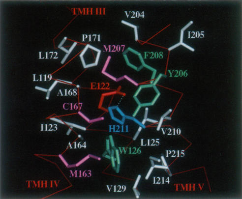
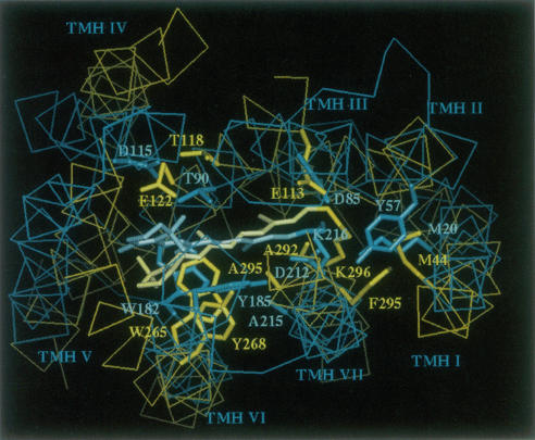
A 3D model of the transmembrane 7-alpha-bundle of rhodopsin-like G-protein-coupled receptors (GPCRs) was calculated using an iterative distance geometry refinement with an evolving system of hydrogen bonds, formed by intramembrane polar side chains in various proteins of the family and collectively applied as distance constraints. The alpha-bundle structure thus obtained provides H bonding of nearly all buried polar side chains simultaneously in the 410 GPCRs considered. Forty evolutionarily conserved GPCR residues form a single continuous domain, with an aliphatic "core" surrounded by six clusters of polar and aromatic side chains. The 7-alpha-bundle of a specific GPCR can be calculated using its own set of H bonds as distance constraints and the common "average" model to restrain positions of the helices. The bovine rhodopsin model thus determined is closely packed, but has a few small polar cavities, presumably filled by water, and has a binding pocket that is complementary to 11-cis (6-s-cis, 12-s-trans, C = N anti)-retinal or to all-trans-retinal, depending on conformations of the Lys296 and Trp265 side chains. A suggested mechanism of rhodopsin photoactivation, triggered by the cis-trans isomerization of retinal, involves rotations of Glu134, Tyr223, Trp265, Lys296, and Tyr306 side chains and rearrangement of their H bonds. The model is in agreement with published electron cryomicroscopy, mutagenesis, chemical modification, cross-linking, Fourier transform infrared spectroscopy, Raman spectroscopy, electron paramagnetic resonance spectroscopy, NMR, and optical spectroscopy data. The rhodopsin model and the published structure of bacteriorhodopsin have very similar retinal-binding pockets.

摘要

利用迭代距离几何优化方法,结合由该家族各种蛋白质中的膜内极性侧链形成并共同用作距离约束的不断演化的氢键系统,计算出了视紫红质样G蛋白偶联受体(GPCR)的跨膜7α束三维模型。由此获得的α束结构在考虑的410种GPCR中几乎同时为所有埋藏的极性侧链提供了氢键。40个进化保守的GPCR残基形成一个单一的连续结构域,其脂肪族“核心”被六簇极性和芳香族侧链包围。特定GPCR的7α束可以使用其自身的氢键集作为距离约束,并结合通用的“平均”模型来限制螺旋的位置。由此确定的牛视紫红质模型紧密堆积,但有一些小的极性腔,可能被水填充,并且有一个与11-顺式(6-s-顺式,12-s-反式,C = N反式)视黄醛或全反式视黄醛互补的结合口袋,这取决于Lys296和Trp265侧链的构象。视黄醛的顺反异构化引发视紫红质光激活的一种推测机制涉及Glu134、Tyr223、Trp265、Lys296和Tyr306侧链的旋转及其氢键的重排。该模型与已发表的电子冷冻显微镜、诱变、化学修饰、交联、傅里叶变换红外光谱、拉曼光谱、电子顺磁共振光谱、核磁共振和光学光谱数据一致。视紫红质模型和已发表的细菌视紫红质结构具有非常相似的视黄醛结合口袋。

https://cdn.ncbi.nlm.nih.gov/pmc/blobs/fc9d/1184393/751f6a69ff29/biophysj00034-0070-a.jpg
https://cdn.ncbi.nlm.nih.gov/pmc/blobs/fc9d/1184393/d62286d7d460/biophysj00034-0064-a.jpg
https://cdn.ncbi.nlm.nih.gov/pmc/blobs/fc9d/1184393/2efe2752909c/biophysj00034-0065-a.jpg
https://cdn.ncbi.nlm.nih.gov/pmc/blobs/fc9d/1184393/751f6a69ff29/biophysj00034-0070-a.jpg
https://cdn.ncbi.nlm.nih.gov/pmc/blobs/fc9d/1184393/d62286d7d460/biophysj00034-0064-a.jpg
https://cdn.ncbi.nlm.nih.gov/pmc/blobs/fc9d/1184393/2efe2752909c/biophysj00034-0065-a.jpg
https://cdn.ncbi.nlm.nih.gov/pmc/blobs/fc9d/1184393/751f6a69ff29/biophysj00034-0070-a.jpg

相似文献

1
The transmembrane 7-alpha-bundle of rhodopsin: distance geometry calculations with hydrogen bonding constraints.视紫红质的跨膜7α束:具有氢键约束的距离几何计算
Biophys J. 1997 May;72(5):1963-85. doi: 10.1016/S0006-3495(97)78842-8.
2
Opioid receptor three-dimensional structures from distance geometry calculations with hydrogen bonding constraints.基于氢键约束的距离几何计算得出的阿片受体三维结构。
Biophys J. 1998 Aug;75(2):612-34. doi: 10.1016/S0006-3495(98)77552-6.
3
Agonist-induced conformational changes in bovine rhodopsin: insight into activation of G-protein-coupled receptors.激动剂诱导的牛视紫红质构象变化:深入了解G蛋白偶联受体的激活
J Mol Biol. 2008 Oct 3;382(2):539-55. doi: 10.1016/j.jmb.2008.06.084. Epub 2008 Jul 7.
4
Structure of bovine rhodopsin in a trigonal crystal form.三角晶型牛视紫红质的结构
J Mol Biol. 2004 Nov 5;343(5):1409-38. doi: 10.1016/j.jmb.2004.08.090.
5
Functional role of internal water molecules in rhodopsin revealed by X-ray crystallography.X射线晶体学揭示视紫红质中内部水分子的功能作用。
Proc Natl Acad Sci U S A. 2002 Apr 30;99(9):5982-7. doi: 10.1073/pnas.082666399. Epub 2002 Apr 23.
6
Specific tryptophan UV-absorbance changes are probes of the transition of rhodopsin to its active state.特定的色氨酸紫外线吸收变化是视紫红质向其活性状态转变的探针。
Biochemistry. 1996 Aug 27;35(34):11149-59. doi: 10.1021/bi960858u.
7
A ligand channel through the G protein coupled receptor opsin.一种通过G蛋白偶联受体视蛋白的配体通道。
PLoS One. 2009;4(2):e4382. doi: 10.1371/journal.pone.0004382. Epub 2009 Feb 5.
8
The steric trigger in rhodopsin activation.视紫红质激活中的空间触发因素。
J Mol Biol. 1997 Jun 13;269(3):373-84. doi: 10.1006/jmbi.1997.1035.
9
Functional role of the "ionic lock"--an interhelical hydrogen-bond network in family A heptahelical receptors.“A类七螺旋受体中螺旋间氢键网络‘离子锁’的功能作用”
J Mol Biol. 2008 Jul 18;380(4):648-55. doi: 10.1016/j.jmb.2008.05.022. Epub 2008 May 17.
10
Complex of an active mu-opioid receptor with a cyclic peptide agonist modeled from experimental constraints.基于实验限制构建的活性μ阿片受体与环肽激动剂的复合物。
Biochemistry. 2004 Dec 21;43(50):15796-810. doi: 10.1021/bi048413q.

引用本文的文献

1
Molecular Mechanisms Underlying the Spectral Shift in Zebrafish Cone Opsins.斑马鱼视锥视蛋白光谱位移的分子机制
bioRxiv. 2024 Sep 24:2024.09.24.614827. doi: 10.1101/2024.09.24.614827.
2
Translation of structure-activity relationships from cyclic mixed efficacy opioid peptides to linear analogues.从环状混合效价阿片肽到线性类似物的构效关系翻译
Biopolymers. 2014 Jan;102(1):107-14. doi: 10.1002/bip.22437.
3
Lipid receptor S1P₁ activation scheme concluded from microsecond all-atom molecular dynamics simulations.从微秒全原子分子动力学模拟中得出的脂质受体 S1P₁激活方案。

本文引用的文献

1
In situ microspectrophotometric studies on the pigments of single retinal rods.单个视杆细胞色素的原位显微分光光度研究。
Biophys J. 1962 Mar;2(2 Pt 1):161-78. doi: 10.1016/s0006-3495(62)86847-7.
2
TAUTOMERIC FORMS OF METARHODOPSIN.变视紫红质的互变异构形式
J Gen Physiol. 1963 Nov;47(2):215-40. doi: 10.1085/jgp.47.2.215.
3
Functional interaction of transmembrane helices 3 and 6 in rhodopsin. Replacement of phenylalanine 261 by alanine causes reversion of phenotype of a glycine 121 replacement mutant.视紫红质中跨膜螺旋3和6的功能相互作用。将苯丙氨酸261替换为丙氨酸会导致甘氨酸121替换突变体表型的逆转。
PLoS Comput Biol. 2013;9(10):e1003261. doi: 10.1371/journal.pcbi.1003261. Epub 2013 Oct 3.
4
Unique mechanisms of excitation energy transfer, electron transfer and photoisomerization in biological systems.生物系统中激发能转移、电子转移和光异构化的独特机制。
J Biol Phys. 2002 Sep;28(3):367-81. doi: 10.1023/A:1020356404203.
5
Modulation of opioid receptor ligand affinity and efficacy using active and inactive state receptor models.利用激活态和失活态受体模型调节阿片受体配体亲和力和效能。
Chem Biol Drug Des. 2012 Nov;80(5):763-70. doi: 10.1111/cbdd.12014. Epub 2012 Sep 12.
6
FoldGPCR: structure prediction protocol for the transmembrane domain of G protein-coupled receptors from class A.FoldGPCR:A 类 G 蛋白偶联受体跨膜结构域的结构预测方案。
Proteins. 2010 Aug 1;78(10):2189-201. doi: 10.1002/prot.22731.
7
Pentapeptides displaying mu opioid receptor agonist and delta opioid receptor partial agonist/antagonist properties.具有μ阿片受体激动剂和δ阿片受体部分激动剂/拮抗剂特性的五肽。
J Med Chem. 2009 Dec 10;52(23):7724-31. doi: 10.1021/jm9007483.
8
Docking studies suggest ligand-specific delta-opioid receptor conformations.对接研究表明存在配体特异性的δ阿片受体构象。
J Mol Model. 2009 Mar;15(3):267-80. doi: 10.1007/s00894-008-0396-7. Epub 2008 Dec 4.
9
Homology modeling of opioid receptor-ligand complexes using experimental constraints.利用实验约束条件对阿片受体-配体复合物进行同源建模。
AAPS J. 2005 Oct 5;7(2):E434-48. doi: 10.1208/aapsj070243.
10
A strategy using NMR peptide structures of thromboxane A2 receptor as templates to construct ligand-recognition pocket of prostacyclin receptor.一种以血栓素A2受体的核磁共振肽结构为模板构建前列环素受体配体识别口袋的策略。
BMC Biochem. 2005 Nov 4;6:23. doi: 10.1186/1471-2091-6-23.
J Biol Chem. 1996 Dec 13;271(50):32337-42. doi: 10.1074/jbc.271.50.32337.
4
Structural features and light-dependent changes in the cytoplasmic interhelical E-F loop region of rhodopsin: a site-directed spin-labeling study.视紫红质细胞质中螺旋间E-F环区域的结构特征及光依赖性变化:一项定点自旋标记研究
Biochemistry. 1996 Sep 24;35(38):12470-8. doi: 10.1021/bi960849l.
5
Structural classification of proteins: new superfamilies.蛋白质的结构分类:新的超家族
Curr Opin Struct Biol. 1996 Jun;6(3):386-94. doi: 10.1016/s0959-440x(96)80059-5.
6
Activating mutations of rhodopsin and other G protein-coupled receptors.视紫红质及其他G蛋白偶联受体的激活突变
Annu Rev Biophys Biomol Struct. 1996;25:287-314. doi: 10.1146/annurev.bb.25.060196.001443.
7
Specific tryptophan UV-absorbance changes are probes of the transition of rhodopsin to its active state.特定的色氨酸紫外线吸收变化是视紫红质向其活性状态转变的探针。
Biochemistry. 1996 Aug 27;35(34):11149-59. doi: 10.1021/bi960858u.
8
Projection structure of an invertebrate rhodopsin.一种无脊椎动物视紫红质的投射结构。
J Struct Biol. 1996 Jul-Aug;117(1):36-44. doi: 10.1006/jsbi.1996.0067.
9
Electron-crystallographic refinement of the structure of bacteriorhodopsin.细菌视紫红质结构的电子晶体学精修
J Mol Biol. 1996 Jun 14;259(3):393-421. doi: 10.1006/jmbi.1996.0328.
10
Identification of amino acid residues in transmembrane helices VI and VII of the lutropin/choriogonadotropin receptor involved in signaling.鉴定促黄体生成素/绒毛膜促性腺激素受体跨膜螺旋VI和VII中参与信号传导的氨基酸残基。
Biochemistry. 1996 Apr 2;35(13):3986-93. doi: 10.1021/bi952421c.