Newbury Dale E
National Institute of Standards and Technology, Gaithersburg, MD 20899-8371.
J Res Natl Inst Stand Technol. 2002 Dec 1;107(6):567-603. doi: 10.6028/jres.107.048. Print 2002 Nov-Dec.
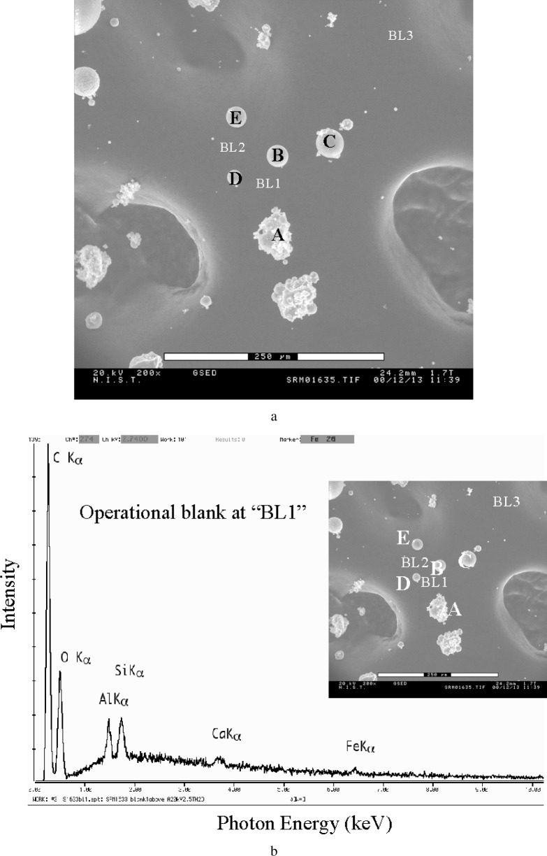
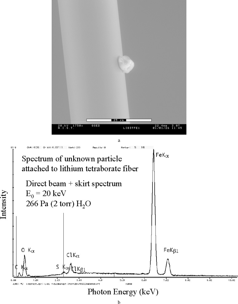
Electron-excited x-ray microanalysis performed in the variable pressure and environmental scanning electron microscopes is subject to additional artifacts beyond those encountered in the conventional scanning electron microscope. Gas scattering leads to direct contributions to the spectrum from the environmental gas, as well as remote generation of x rays by electrons scattered out of the focussed beam. The analyst can exert some degree of control over these artifacts, but depending on the exact situation, spurious elements can appear at the trace (< 0.01 mass fraction), minor (0.01 mass fraction to 0.1 mass fraction), or even major (> 0.1 mass fraction) levels. Dispersed particle samples give the least compromised results, while fine scale microstructures are the most severely compromised. Procedures to optimize the situation based upon specimen preparation as well as spectral processing are described.
在可变压力和环境扫描电子显微镜中进行的电子激发X射线微分析,除了传统扫描电子显微镜中遇到的伪像外,还会受到其他伪像的影响。气体散射会导致环境气体直接对光谱产生贡献,以及从聚焦束中散射出来的电子远程产生X射线。分析人员可以对这些伪像进行一定程度的控制,但根据具体情况,杂质元素可能会以痕量(<0.01质量分数)、微量(0.01质量分数至0.1质量分数)甚至主量(>0.1质量分数)水平出现。分散颗粒样品得到的结果受影响最小,而精细尺度的微观结构受影响最严重。本文描述了基于样品制备和光谱处理来优化这种情况的程序。