Xu Xiaowen, Wang Liang, Chen Liying, Su Tianjiao, Zhang Yan, Wang Tiantian, Ma Weifeng, Yang Fan, Zhai Wujie, Xie Yuanyuan, Li Dan, Chen Qiong, Fu Xuemei, Ma Yuanzheng, Zhang Yan
Center of Orthopedics, The 309th Hospital of PLA, Beijing, 100091, China.
Center for Systems Biomedical Sciences, University of Shanghai for Science and Technology, Shanghai, 200093, China.
J Orthop Surg Res. 2016 Aug 2;11(1):87. doi: 10.1186/s13018-016-0418-6.
This study aimed to assess the effects of chronic sleep deprivation (CSD) on bone mass and bone metabolism in rats.
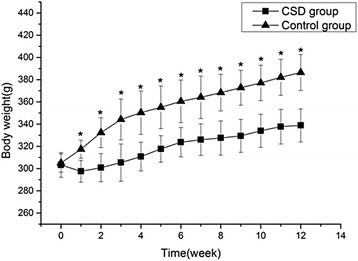
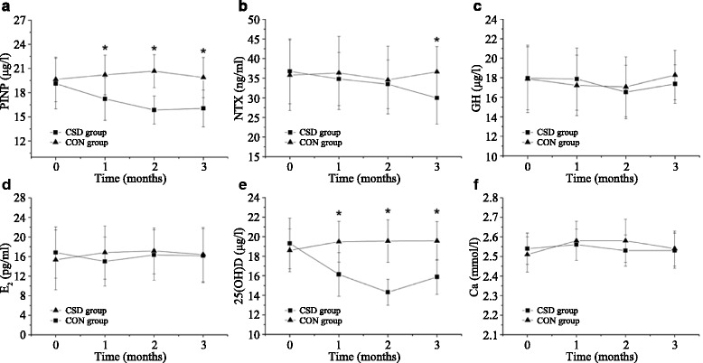
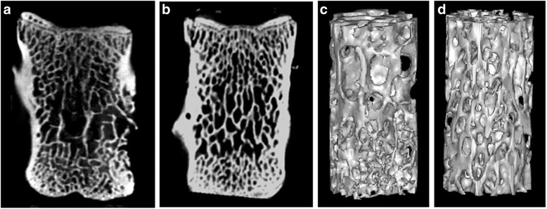
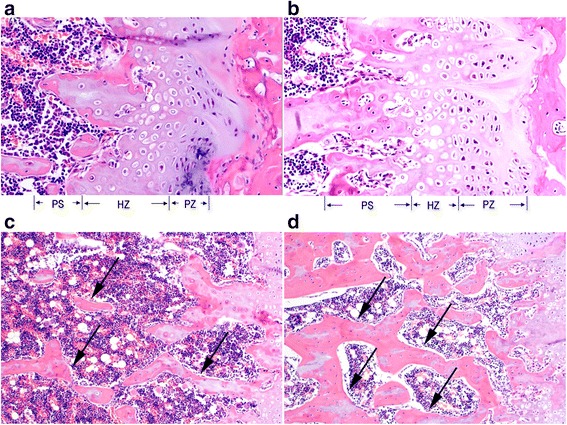
Twenty-four rats were randomly divided into CSD and control (CON) groups. Rats were subjected to CSD by using the modified multiple platform method (MMPM) to establish an animal model of CSD. Biochemical parameters such as levels of serum N-terminal propeptide of type I procollagen (PINP), N-terminal cross-linking telopeptide of type I collagen (NTX), growth hormone (GH), estradiol (E2), serum 25(OH)D, and calcium (Ca) were evaluated at 0, 1, 2, and 3 months. After 3 months, each fourth lumbar vertebra and the distal femoral metaphysis of the left extremity of rats were harvested for micro-computed tomography scans and histological analysis, respectively, after the rats were sacrificed under an overdose of pentobarbital sodium.
Compared with rats from the CON group, rats from the CSD group showed significant decreases in bone mineral density (BMD), bone volume over total volume, trabecular bone thickness, and trabecular bone number and significant increases in bone surface area over bone volume and trabecular bone separations (P < 0.05). Bone histomorphology studies showed that rats in the CSD group had decreased osteogenesis, impaired mineralization of newly formed bones, and deteriorative trabecular bone in the secondary spongiosa zone. In addition, they showed significantly decreased levels of serum PINP (1 month later) and NTX (3 months later) (P < 0.05). The serum 25(OH)D level of rats from the CSD group was lower than that of rats from the CON group after 1 month (P < 0.05).
CSD markedly affects bone health by decreasing BMD and 25(OH)D, deteriorating the bone microarchitecture, and decreasing bone formation and bone resorption markers.
本研究旨在评估慢性睡眠剥夺(CSD)对大鼠骨量和骨代谢的影响。
将24只大鼠随机分为CSD组和对照组(CON)。采用改良多平台法(MMPM)使大鼠遭受慢性睡眠剥夺,以建立CSD动物模型。在0、1、2和3个月时评估血清I型前胶原N端前肽(PINP)、I型胶原N端交联端肽(NTX)、生长激素(GH)、雌二醇(E2)、血清25(OH)D和钙(Ca)等生化参数。3个月后,在大鼠过量戊巴比妥钠麻醉下处死后,分别采集每只大鼠的第四腰椎和左下肢股骨远端干骺端进行微计算机断层扫描和组织学分析。
与CON组大鼠相比,CSD组大鼠的骨矿物质密度(BMD)、骨体积与总体积之比、小梁骨厚度和小梁骨数量显著降低,骨表面积与骨体积之比和小梁骨间距显著增加(P < 0.05)。骨组织形态学研究表明,CSD组大鼠成骨减少,新形成骨矿化受损,次级海绵骨区小梁骨恶化。此外,它们的血清PINP水平(1个月后)和NTX水平(3个月后)显著降低(P < 0.05)。1个月后,CSD组大鼠的血清25(OH)D水平低于CON组大鼠(P < 0.05)。
慢性睡眠剥夺通过降低骨密度和25(OH)D、恶化骨微结构以及降低骨形成和骨吸收标志物,显著影响骨骼健康。