Christidi Foteini, Karavasilis Efstratios, Samiotis Kostantinos, Bisdas Sotirios, Papanikolaou Nikolaos
1st Department of Neurology, Aeginition Hospital, Medical School, National and Kapodistrian University, Athens, Greece.
2nd Department of Radiology, University General Hospital 'Attikon', School of Medicine, National and Kapodistrian University of Athens, Athens, Greece.
Eur J Radiol Open. 2016 Jul 18;3:153-61. doi: 10.1016/j.ejro.2016.06.002. eCollection 2016.
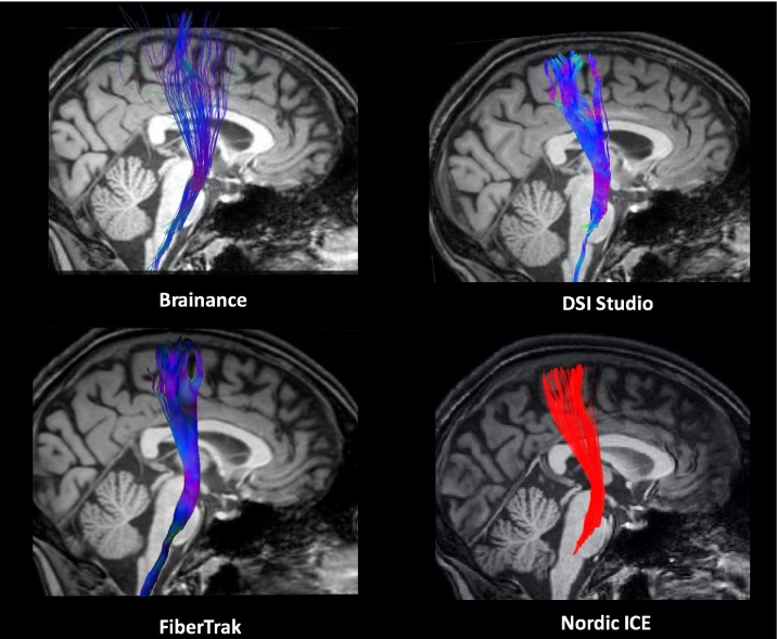
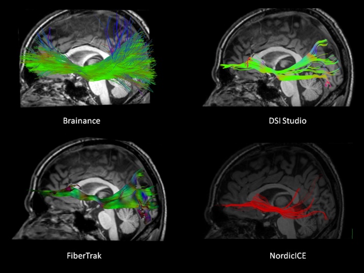
Diffusion tensor imaging (DTI) enables in vivo reconstruction of white matter (WM) pathways. Considering the emergence of numerous models and fiber tracking techniques, we herein aimed to compare, both quantitatively and qualitatively, the fiber tracking results of four DTI software (Brainance, Philips FiberTrak, DSI Studio, NordicICE) on the reconstruction of representative WM tracts.
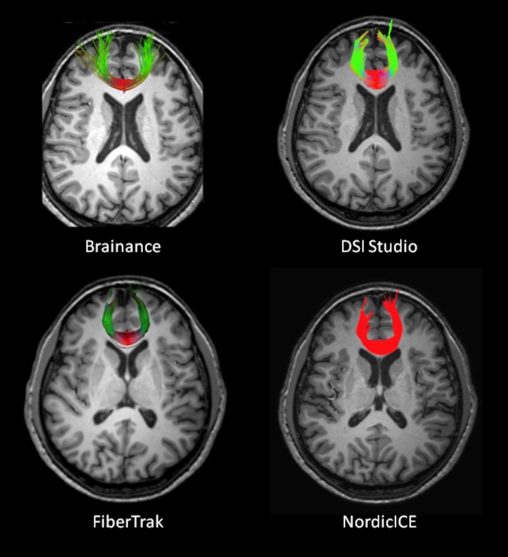
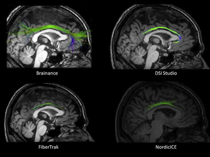
Ten healthy participants underwent 30-directional diffusion tensor imaging on a 3T-Philips Achieva TX MR-scanner. All data were analyzed by two independent sites of experienced raters with the aforementioned software and the following WM tracts were reconstructed: corticospinal tract (CST); forceps major (Fmajor); forceps minor (Fminor); cingulum bundle (CB); superior longitudinal fasciculus (SLF); inferior fronto-occipital fasciculus (IFOF). Visual inspection of the resulted tracts and statistical analysis (inter-rater and betweensoftware agreement; paired t-test) on fractional anisotropy (FA), axial and radial diffusivity (Daxial, Dradial) were applied for qualitative and quantitative evaluation of DTI software results.
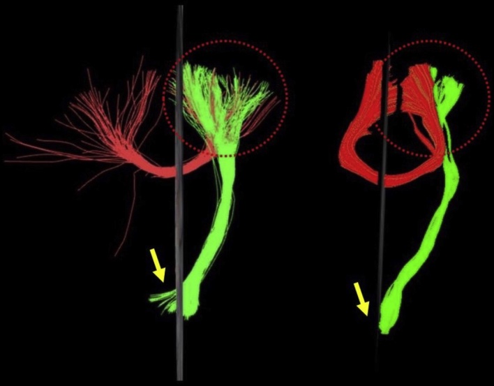
Qualitative evaluation of the extracted tracts confirmed anatomical landmarks at least for the core part of each tract, even though differences in the number of fibers extracted and the whole tract were evident, especially for the CST, Fmajor, Fminor and SLF. Descriptive values did not deviate from the expected range of values for healthy adult population. Substantial inter-rater agreement (intraclass correlation coefficient [ICC], Bland-Altman analysis) was found for all tracts (ICC; FA: 0.839-0.989, Daxial: 0.704-0.991, Dradial: 0.972-0.993). Low agreement for FA, Daxial and Dradial (ICC; Bland-Altman analysis) and significant paired t-test differences (p < 0.05) were detected regarding between-software agreement.
Qualitative comparison of four different DTI software in addition to substantial inter-rater but poor between-software agreement highlight the differences on existing fiber tracking methodologies and several particularities of each WM tract, further supporting the need for further study in both clinical and research settings.
扩散张量成像(DTI)能够在体内重建白质(WM)通路。鉴于众多模型和纤维追踪技术的出现,我们在此旨在对四种DTI软件(Brainance、飞利浦纤维追踪、DSI Studio、NordicICE)在重建代表性WM束方面的纤维追踪结果进行定量和定性比较。
10名健康受试者在3T飞利浦Achieva TX磁共振扫描仪上进行了30方向扩散张量成像。所有数据由两个独立的经验丰富的评估者使用上述软件进行分析,并重建以下WM束:皮质脊髓束(CST);主要钳状束(Fmajor);次要钳状束(Fminor);扣带束(CB);上纵束(SLF);额枕下束(IFOF)。对所得束进行视觉检查,并对分数各向异性(FA)、轴向和径向扩散率(Daxial、Dradial)进行统计分析(评估者间和软件间一致性;配对t检验),以对DTI软件结果进行定性和定量评估。
对提取束的定性评估至少在每条束的核心部分确认了解剖标志,尽管提取的纤维数量和整个束存在明显差异,特别是对于CST、Fmajor、Fminor和SLF。描述性值未偏离健康成年人群的预期值范围。所有束均发现评估者间具有高度一致性(组内相关系数[ICC],Bland-Altman分析)(ICC;FA:0.839 - 0.989,Daxial:0.704 - 0.991,Dradial:0.972 - 0.993)。在软件间一致性方面,检测到FA、Daxial和Dradial的一致性较低(ICC;Bland-Altman分析)以及显著的配对t检验差异(p < 0.05)。
除了评估者间高度一致但软件间一致性较差外,对四种不同DTI软件的定性比较突出了现有纤维追踪方法的差异以及每条WM束的几个特点,进一步支持了在临床和研究环境中进行进一步研究的必要性。