Univ. de Bordeaux, Institut des Maladies Neurodégénératives, UMR 5293, F-33000 Bordeaux, France.
CNRS, Institut des Maladies Neurodégénératives, UMR 5293, F-33000 Bordeaux, France.
Sci Rep. 2016 Sep 14;6:31836. doi: 10.1038/srep31836.
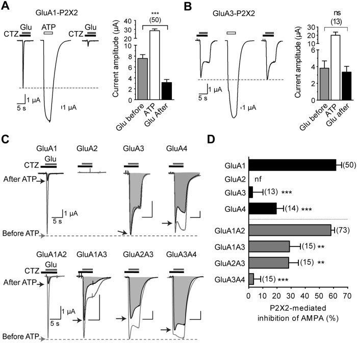
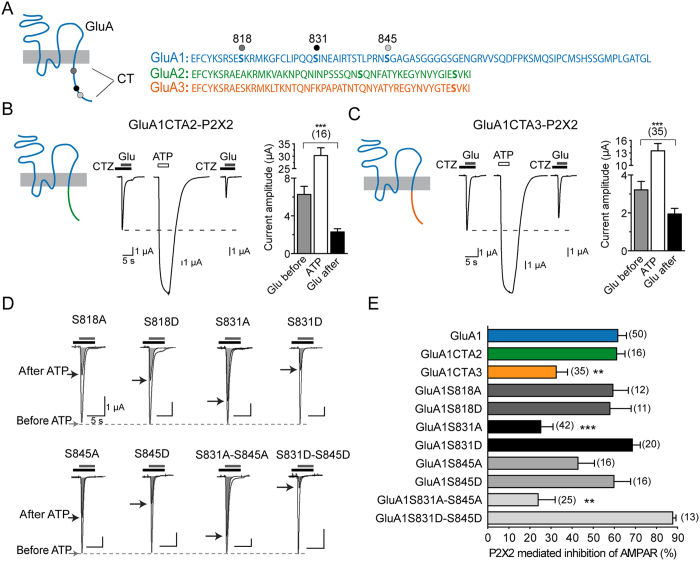
Plasticity at excitatory synapses can be induced either by synaptic release of glutamate or the release of gliotransmitters such as ATP. Recently, we showed that postsynaptic P2X2 receptors activated by ATP released from astrocytes downregulate synaptic AMPAR, providing a novel mechanism by which glial cells modulate synaptic activity. ATP- and lNMDA-induced depression in the CA1 region of the hippocampus are additive, suggesting distinct molecular pathways. AMPARs are homo-or hetero-tetramers composed of GluA1-A4. Here, we first show that P2X2-mediated AMPAR inhibition is dependent on the subunit composition of AMPAR. GluA3 homomers are insensitive and their presence in heteromers alters P2X-mediated inhibition. Using a mutational approach, we demonstrate that the two CaMKII phosphorylation sites S567 and S831 located in the cytoplasmic Loop1 and C-terminal tail of GluA1 subunits, respectively, are critical for P2X2-mediated AMPAR inhibition recorded from co-expressing Xenopus oocytes and removal of surface AMPAR at synapses of hippocampal neurons imaged by the super-resolution dSTORM technique. Finally, using phosphorylation site-specific antibodies, we show that P2X-induced depression in hippocampal slices produces a dephosphorylation of the GluA1 subunit at S567, contrary to NMDAR-mediated LTD. These findings indicate that GluA1 phosphorylation of S567 and S831 is critical for P2X2-mediated AMPAR internalization and ATP-driven synaptic depression.
兴奋性突触的可塑性可以通过谷氨酸盐的突触释放或诸如 ATP 的神经胶质递质的释放来诱导。最近,我们表明,由星形胶质细胞释放的 ATP 激活的突触后 P2X2 受体下调 AMPAR,提供了神经胶质细胞调节突触活性的新机制。ATP 和 lNMDA 在海马 CA1 区诱导的抑制是相加的,表明存在不同的分子途径。AMPA 受体是由 GluA1-A4 组成的同型或异型四聚体。在这里,我们首先表明,P2X2 介导的 AMPAR 抑制依赖于 AMPAR 的亚基组成。GluA3 同型体不敏感,其在异型体中的存在改变了 P2X 介导的抑制。通过突变方法,我们证明位于细胞质环 1 和 GluA1 亚基 C 末端尾部中的两个 CaMKII 磷酸化位点 S567 和 S831 对于从共表达的非洲爪蟾卵母细胞记录的 P2X2 介导的 AMPAR 抑制以及通过超分辨率 dSTORM 技术成像的海马神经元突触处的表面 AMPAR 的去除是至关重要的。最后,使用磷酸化位点特异性抗体,我们表明,与 NMDAR 介导的 LTD 相反,海马切片中的 P2X 诱导的抑制导致 S567 处的 GluA1 亚基去磷酸化。这些发现表明,GluA1 亚基 S567 和 S831 的磷酸化对于 P2X2 介导的 AMPAR 内化和 ATP 驱动的突触抑制至关重要。